AUTHOR OF THIS BLOG

DR ANTHONY MELVIN CRASTO, WORLDDRUGTRACKER

DR ANTHONY MELVIN CRASTO Ph.D

DR ANTHONY MELVIN CRASTO, Worlddrugtracker, Born in Mumbai in 1964 and graduated from Mumbai University, Completed his PhD from ICT ,1991, Mumbai, India, in Organic chemistry, The thesis topic was Synthesis of Novel Pyrethroid Analogues, Currently he is working with AFRICURE PHARMA as ADVISOR earlier GLENMARK LS Research centre as consultant,Principal Scientist, Process Research (bulk actives) at Mahape, Navi Mumbai, India. Prior to joining Glenmark, he worked with major multinationals like Hoechst Marion Roussel, now sSanofi, Searle India ltd, now Rpg lifesciences, etc. he is now helping millions, has million hits on google on all organic chemistry websites. His New Drug Approvals, Green Chemistry International, Eurekamoments in organic chemistry are some most read blogs He has hands on experience in initiation and developing novel routes for drug molecules and implementation them on commercial scale over a 32 year tenure, good knowledge of IPM, GMP, Regulatory aspects, he has several international drug patents published worldwide . He gas good proficiency in Technology transfer, Spectroscopy, Stereochemistry, Synthesis, polymorphism etc He suffered a paralytic stroke in dec 2007 and is bound to a wheelchair, this seems to have injected feul in him to help chemists around the world, he is more active than before and is pushing boundaries, he has one lakh connections on all networking sites, He makes himself available to all, contact him on +91 9323115463, [email protected]

2-Hydroxymalonitrile-A Useful Reagent for One-step Synthesis of α-Hydroxy Esters

 spectroscopy, SYNTHESIS  Comments Off on 2-Hydroxymalonitrile-A Useful Reagent for One-step Synthesis of α-Hydroxy Esters
Jun 042015


YANG Jianxin, YIN Yunxing, HE Zhenmin, MA Li, LI Xin, ZHANG Zhiliu, LIN Xiaojuan, MA Rujian
2-Hydroxymalonitrile-A Useful Reagent for One-step Synthesis of α-Hydroxy Esters
2015 Vol. 31 (3): 321-324 [Abstract] ( 47 ) [HTML 1KB] [PDF 0KB] ( 68 )
doi10.1007/s40242-015-4495-6

see

http://www.cjcu.jlu.edu.cn/hxyj/EN/abstract/abstract16155.shtml


2-Hydroxymalonitrile-A Useful Reagent for One-step Synthesis of α-Hydroxy Esters
YANG Jianxin1,2, YIN Yunxing2, HE Zhenmin2, MA Li2, LI Xin2, ZHANG Zhiliu2, LIN Xiaojuan2, MA Rujian2
1. Tianjin Key Laboratory for Modern Drug Delivery & High-Efficiency, School of Pharmaceutical Science and Technology, Tianjin University, Tianjin 300072, P. R. China;
2. WuXi PharmaTech Co. Ltd., Shanghai 200131, P. R. China
Corresponding Authors: MA Rujian     E-mail: [email protected]


TAKE A TOUR


Tianjin china

Map of tianjin

Image result for tianjin china map.


.


/////////

Share

KETO ENOL TAUTOMERISM AND NMR

 spectroscopy, Uncategorized  Comments Off on KETO ENOL TAUTOMERISM AND NMR
Jun 032015
 

.


H Nmr Spectrum | Apk Mod Game

www.apkmodgame.net

Shows a method for getting all the useful information out of a proton nmr spectrum and using it to piece together the identity of an unknown molecule.
A Partial NMR Spectrum of 2,4-Pentanedione


Patent EP0922715B1 – Stimuli-responsive polymer utilizing keto …

Carbonyl compounds (aldehydes, ketones, carboxylic esters, carboxylic amides) react aselectrophiles at the sp2 hybridized carbon atoms and as nucleophiles if they contain an H-atom in the α-position relative to their C=O or C=N bonds. This is because this H is acidic and it can be removed by a base leaving behind an electron pair for nucleophilic attacks.

For most compounds in organic chemistry all the molecules have the same structure – even if this structure cannot satisfactory represented by a Lewis formula – but for many compounds there is a mixture of two or more structurally distinct compounds that are in rapid equilibrium. This phenomenon is called tautomerism.

Tautomerism is the phenomenon that occurs in any reaction that simply involves the intramolecular transfer of a proton. An equilibrium is established between the two tautomers (structurally distinct compounds) and there is a rapid shift back and forth between the distinct compounds.

A very common form of tautomerism is that between a carbonyl compound containing an αhydrogen and its enol form (Fig. I.1).

Fig. I.1: A keto-enol reaction


An enol is exactly what the name implies: an ene-ol. It has a C=C double bond (diene) and an OH group (alcohol) joined directly to it.

Notice that in the above reaction as in any keto-enol reaction there is no change in pH since a proton is lost from carbon and gained on oxygen. The reaction is known as enolization as it is the conversion of a carbonyl compound into its enol.

Notice also that in the above reaction the product is almost the same as the starting material since the only change is the transfer of one proton and the shift of the double bond.

In simple cases (R2 = H, alkyl, OR, etc.) the equilibrium of the keto-enol reaction lies well to the left (keto structure) (Table I.1). The reason can be seen by examining the bond energies in Table I.2.


Compound

Enol Content, %

Acetone

6 * 10-7

PhCOCH3

1.1 * 10-6

CH3CHO

6 * 10-5

Cyclohexanone

4 * 10-5

Ph2CHCHO

9.1

PhCOCH2COCH3

89.2

Table I.1: The enol content of some carbonyl compounds


If keto-enol reactions are common for aldehydes and ketones why don’t simple aldehydes and ketones exist as enols?

IR and NMR Spectra of carbonyl compounds show no signs of enols. The equilibrium lies well over towards the keto form (the equilibrium constant k for cyclohexanone is about 10-5).


Bond (Energy, kJ/mol)

Sum ( kJ/mol)

keto form

C-H (413)

C-C (350)

C=O (740)

1503

enol form

C=C (620)

C-O (367)

O-H (462)

1449

Table I.2: Bond energies in the keto and in the enol form. The keto form is thermodynamically more stable than the enol form by approximately 50 kJ/mol

The approximate sum of the bond energies in the keto form is 1503 kJ/mol while in the enol form 1449. Therefore, the keto form is thermodynamically more stable than the enol form by approximately 50 kJ/mol.

In most cases, enol forms cannot be isolated since they are less stable and are formed in minute quantities. However, there are some exceptions and in certain cases a larger amount of the enol form is present and it can be even the predominant species:

  • Molecules in which the enolic double bond is in conjugation with another double bond (cases are shown in Table I.1 like Ph2CHCHO and PhCOCH2COCH3)
  • Molecules that contain two or more bulky aryl groups (Fig. I.2). Compound I in Fig. I.2 (a substituted enol) is the major species in equilibrium (~95%) while the keto form is the minor species (~5%). In cases like this steric hindrance destabilizes the keto form (the two substituted aryl groups are 109° apart) while in the enol form 120° apart.


Fig. I.2: A keto-enol reaction. The enol form (I) is the major species since the keto form is destabilized by steric hindrance (the substituted aryl groups are closer in the keto form – the C-C angle is 109° and this is unfavorable due to steric hindrance)
Fig. I.2: A keto-enol reaction. The enol form (I) is the major species in this case since the keto form is destabilized by steric hindrance (the substituted aryl groups are closer in the keto form – the C-C angle is 109° and this is unfavorable due to steric hindrance)


Is there experimental evidence that keto-enol reactions are common for aldehydes and ketones?

If the NMR spectrum of a simple carbonyl compound in D2O is obtained – such as pinacolone’s (CH3)3CCOCH3 – the signal for protons next to the carbonyl group very slowly disappears. The isolated compound’s mass spectrum (after the above mentioned reaction with D2O is over) shows that those hydrogen atoms have been replaced by deuterium atoms. There is a peak at (M+1)+ or (M+2)+ or (M+3)+ instead of M+. The reaction is shown in Fig. I.3:

 Fig. I.3: Evidence for a keto-enol reaction when pinacolone (CH3)3CCOCH3 reacts with D2O. When the enol form of the pinacolone reverts to the keto form it picks up a deuteron instead of a proton because the solution consists almost entirely of D2O.
Fig. I.3: Evidence for a keto-enol reaction when pinacolone (CH3)3CCOCH3 reacts with D2O. When the enol form of the pinacolone reverts to the keto form it picks up a deuteron instead of a proton because the solution consists almost entirely of D2O.


What mechanism can be proposed for the above reaction?

Enolization is a slow process in neutral solution, even in D2O, and is catalyzed by acid or base in order to happen.

In the acid-catalyzed reaction the molecule is first protonated on oxygen and then loses the C-H proton in a second step (Fig. I.4). When the enol form reverts to the keto – since this is an equilibrium process – it picks up a deuteron instead of a proton since the solution is D2O.


Fig. I.4: The acid-catalyzed keto-enol reaction mechanism. If D2O is the solvent then the α-hydrogens to carbonyl group are replaced by deuterium.
Fig. I.4: The acid-catalyzed keto-enol reaction mechanism. If D2O is the solvent then the α-hydrogens to carbonyl group are replaced by deuterium.

In the base-catalyzed reaction the C-H proton is removed first by the base (for example hydroxide ion OH, OD in our case) and the proton (or D+ in our case) added to the oxygen atom in a second step (Fig. I.5).

Fig. I.5: The base-catalyzed keto-enol reaction mechanism. If D2O is the solvent then the α-hydrogens to carbonyl group are replaced by deuterium.
Fig. I.5: The base-catalyzed keto-enol reaction mechanism. If D2O is the solvent then the α-hydrogens to carbonyl group are replaced by deuterium.

Notice that the enolization reactions in Fig. I.4 and Fig. I.5 are catalytic. In the acid-catalyzed mechanism the D+ (or H+ if water is the solvent) is regenerated at the end (catalyst). In the base-catalyzed mechanism OD (or OH if water is the solvent) is regenerated at the end (catalyst).

The enolate ion generated from the enol (Fig. I.6) in the base-catalyzed mechanism is nucleophilic due to:

  • Oxygen’s small atomic radius
  • Formal negative charge

An enolate ion is an ion with a negative charge on oxygen with adjacent C-C double bond.


 Fig. I.6: Enolate ion resonance contributors. Although the major contributor is resonace structure I when it reacts as a nucleophile structure II is more reactive.
Fig. I.6: Enolate ion resonance contributors. Although the major contributor is resonace structure I when it reacts as a nucleophile structure II is more reactive.

Enolates are reactive nucleophiles. Although the major enolate Lewis contributor shows concentration of electron density on the electronegative oxygen when it reacts as a nucleophile, it behaves like the electron density is concentrated on the α-carbon next to carbonyl group.

Enolates react with alkyl halides, aldehydes/ketones and esters and these reactions are shown in the post entitled “The chemistry of enolate ions – Enolate ion reactions”.


References
  1. A.J. Kresge, Pure Appl. Chem., 63, 213 (1991)
  2. B. Capon, The Chemistry of Enols, Wiley, NY, 307–322 (1990)
  3. S.E. Biali et al., J. Am. Chem. Soc. 107, 1007 (1985).


.

http://www.slideshare.net/chemsant/nmr-dynamic


http://article.sapub.org/10.5923.j.ajoc.20140401.01.html

2-fluoro-3-hydroxycyclopent-2-enone and 2-fluoro- 1,3-cyclopentanedione (1c): This compound was obtained as a 52:48 mixture of keto-enol and diketo tautomers in 50% yield as a yellow-brown solid, mp 70-72°C. NMR:1H: δ 2.36 (t, 3JH-H = 16.2 Hz, 2H), 2.85 (m, 2H), 5.91 (d, 2JH-F = 47.7 Hz, 1H). 13C: δ31.1, 90.8 (d, 1JC-F = 251.3 Hz), 122.3 (d, 1JC-F = 233.9 Hz), 210.1 (d, 2JC-F = 31.0 Hz). 19F: keto-enol: δ-161.4 (s, 1F); diketo: δ-195.5 (d, 2JF-H = 47.7 Hz, 1F). Analysis calcd for C5H5FO2: C, 51.73, H, 4.34. Found: C, 51.48, H, 4.31.


Take a tour

Surat gujarat india


Map of surat city.

.

ISKCON

Surat-European tombs


Kathiyavadi food, Garden Restaurant, Restaurant in surat, Restaurant, Restaurant Services, Food


/////////

Share

A Simple Organocatalytic Enantioselective Synthesis of Pregabalin

 Uncategorized  Comments Off on A Simple Organocatalytic Enantioselective Synthesis of Pregabalin
Jun 022015
 

Bassas, O.; Huuskonen, J.; Rissanen, K.; Koskinen, A.M.P. ’A Simple Organocatalytic Enantioselective Synthesis of Pregabalin.’ Eur. J. Org. Chem. 2009, 1340-1351.


This paper describes a new procedure for the enantioselective synthesis of the important anticonvulsant drug Pregabalin, which shows biological properties as the (S) enantiomer only. The key step of the synthetic sequence is the Michael addition reaction of Meldrum’s acid to a nitroalkene mediated by a quinidine derived thiourea. A variety of novel catalysts bearing different groups at the thiourea moiety were synthesized and tested. The most successful catalyst that incorporates a trityl substituent provided up to 75 % ee of (S)-4. The conjugate addition reaction was carried out on a multigram scale with low loadings of catalyst (10 mol-%). Moreover, the catalyst can be recycled showing the same capability in chemical yield and asymmetric induction. Then, hydrogenation of nitroalkane 4 followed by decarboxylation of diacid 5 provides Pregabalin hydrochloride in 59 % overall yield. Enantioenrichment by crystallization of the free amino acid 1 improves the (S)/(R) enantiomeric ratio to 9:1.

Author Information

  1. 1Department of Chemistry, Helsinki University of Technology, P. O. Box 6100, 02015 TKK, Espoo, Finland, Fax: +358-94512538
  2. 2NanoScience Center, Department of Chemistry, University of Jyväskylä, P. O. Box 35, 40014 Jyväskylä, Finland
  1. X-ray crystallography.

Email: Ari M. P. Koskinen ([email protected])

http://onlinelibrary.wiley.com/doi/10.1002/ejoc.200801220/abstract


Jyväskylä, Finland

TAKE A TOUR


Jyväskylä, Finland


  1. Jyväskylä – Wikipedia, the free encyclopedia

    en.wikipedia.org/wiki/Jyväskylä

    Jyväskylä (Finnish pronunciation: [ˈjyvæsˌkylæ]) is a city and municipality in CentralFinland in the western part of the Finnish Lakeland. It is the largest city in  …

    History – ‎Etymology – ‎Government – ‎Geography


///////////

Share

Women in Organic Chemistry

 Uncategorized  Comments Off on Women in Organic Chemistry
Jun 012015


List of female scientists before the 21st century – Wikipedia, the …

New

Women in Chemistry Win Student Service Award – News

cns.utexas.edu1667 × 1111Search by image

When Women in Chemistry (WIC), which has been awarded this year’s College of Natural Sciences Student Service Award, set out to survey chemistry graduate …

New

Prof.Vandana Patravale Keynote Speaker at nanobio Australia 2014 …

www.udctalumni.org.in

Vandana Patravale met their VC and Deans along with business tycoons in a special dinner party hosted by AIBC and they were very keen to work with Institute …

New

Prajakta Dandekar | LinkedIn

in.linkedin.com

 Prajakta Dandekar, UGC Assistant Professor in Engineering Sciences & Ramanujan Fellow, Institute Of Chemical Technology

New

New

Women in science | News Center | Wake Forest University

news.wfu.edu495 × 265Search by image

Professor Rebecca Alexander with Ph.D. student Veronica Casina: Still dealing with the

New

Dr Hina Siddiqui, Pakistani scientists, Eurasia Chemistry award
Dr. Hina Siddiqui, a research officer at ICCBS urged women to come towards Science and research.

New

Preshita Desai has received Best Oral Presentation Award …

www.udctalumni.org.in

Preshita Desai has received Best Oral Presentation Award – Newsroom – UDCT Alumni Network. «

New

Dr Deanna D’Alessandro wins L’Oreal Australia For Women in Science …

… from the School of Chemistry, has won a L’Oréal Australia For Women in Science Fellowship worth $20 000 for her work on metal-organic frameworks that …

New

LSU Tops in the Nation at Awarding Chemistry Ph.D. Degrees to …

www.lsu.edu300 × 200Search by image

BATON ROUGE – LSU is the top university in the nation in granting Ph.D. degrees in chemistry to women and underrepresented minority students, according to a …

New

Roxanne Bales vividly remembers the months she spent at a graduate program in organic chemistry in New England.

New


Ji Qi (GRS’06,’11), a graduate student pursuing a Ph.D. in chemistry, has been awarded a 2006-2007 Novartis Fellowship in Organic Chemistry for Women and …

New


LiveDNA: Publications of Suneela Sunil Dhaneshwar

livedna.org

Suneela Sunil Dhaneshwar,

Suneela Dhaneshwar – YouTube

www.youtube.com

Design and Development of Novel Azo Prodrugs using Various Permutations by Dr.Suneela

New


Chemistry professor Julia Kubanek

New


http://chemists.princeton.edu/knowles/

New


The 2014 winners of the The Elsevier Foundation Awards for Early Career Women Scientists in the Developing World accept their prizes at the annual AAAS …

One Organic Chemist One Day: Five Female Chemists Win TWAS Awardsoneorganichemistoneday.blogspot.com

Five Female Chemists Win TWAS Awards

New


Charusita Chakravarty

Women in Science

www.ias.ac.in1758 × 1995Search by image

Ph.D. (1990, Cambridge), FASc, is a recipient of S S Bhatnagar Award, Swarnajayanti Fellowship of the DST, the Bronze Medal of the Chemical Research Society …

New


.

One Organic Chemist One Day: Dr. Shamsun Nahar Khan

oneorganichemistoneday.blogspot.com

Dr. Shamsun Nahar Khan

New


One Organic Chemist One Day


One Organic Chemist One Day: Vinita Gupta, Group President and CEO …

oneorganichemistoneday.blogspot.com

Vinita Gupta, Group President and CEO at Lupin Pharmaceuticals

New


Annaliese K. Franz | UC Davis Department of Chemistry


One Organic Chemist One Day: Dr. Shamsun Nahar Khan

One Organic Chemist One Day: Prof Zuriati Zakaria…….Organic …

Women in Chemistry — Interview with Yamuna Krishnan :: ChemViews …

Women in Chemistry — Interview with Supawan Tantayanon …

Women in Chemistry — Interview with Zeinab Shaaban Abd El-Ati Abou …

Emma Carr, pioneering Physical Organic Chemist | Mount Holyoke College

Stanford Women in STEM Symposium

wiss.stanford.edu

Megan McClory is a lecturer in the Department of Chemistry. She is currently developing

New

Dr. Donna Nelson: Organic Chemist Synthesizing Research …

10 Indian women scientists you should be proud of

www.thealternative.in

She was an organic chemist from India who was known for her work in bio-organic chemistry, including “pioneering work in protein folding” and “ ..

New

Zakya H. Kafafi – 2007 SPIE Women in Optics Planner

spie.org

Head, Organic Optoelectronics Section, Optical Sciences Division, Naval Research Laboratory Washington, DC, USA

New

Blog Archive » Ms. Akiko Yagi has received the L’Oréal-UNESCO For …

synth.chem.nagoya-u.ac.jp

Ms. Akiko Yagi, Doctoral student in the Group of Prof. Itami has received the L’Oréal-UNESCO For Women in Science Award 2014!

New

Women in Nanoscience Blog

New

cl
June | 2007 | News @ Wesleyan

newsletter.blogs.wesleyan.edu

Andrea Roberts, visiting instructor of chemistry, has introduced “green” techniques into her organic chemistry laboratory sections.

New

Women in Chemistry—A European Journal :: ChemViews Magazine …

www.chemistryviews.org

From left to right: Jessica Nilsson, Francesca Novara, Karen Hindson, Katja Glatz, Rachel McGlue, Anne Deveson, Elisabeth von Roedern, Susan Wilkinson, …

New

One Organic Chemist One Day: Ranjala Ratnayake

oneorganichemistoneday.blogspot.com

Ranjala Ratnayake, Ph.D. (Natural Products Chemistry)

New

Sadhana Sathaye

Sadhana Sathaye

Associate professor of Pharmacy(Pharmacology) at Institute Of Chemical Technology

Lo Schiavo Science | University of San Francisco

blogs.usfca.edu

Tami Spector (on the left), professor of organic chemistry, in a chemistry lab with one of her current students: Amera Al-Faleh, a senior majoring in …

New

Kiran Mazumdar Shaw: (born 23 March 1953) She is the Chairman & Managing Director, Biocon Limited a biotechnology company based at Bangalore. She is on the Forbes list of the world’s 100 most powerful women and in business list on top 50 women released by the Financial Times’. In the year 1978, she started Biocon in the garage of her rented house in Bangalore with a seed capital of Rs. 10,000. Now the net worth of the company is more than $ 900 million. Now Biocon produces drugs for cancer, diabetes and auto-immune diseases. Product pipeline includes world’s first oral insulin, currently undergoing Phase III clinical trials.

Winners of the 2014 Elsevier Foundation Awards for Early Career Women Scientists in Developing Countries: (left to right) Dr. Eqbal Mohammed Abdu Dauqan (Biochemistry - Yemen), Dr. Simone Ann Marie Badal McCreath (Biochemistry - Jamaica), Dr. Taiwo Olayemi Elufioye (Pharmacology - Nigeria), Dr. Leni Ritmaleni (Medicinal Chemistry - Indonesia) and Dr. Nilufar Mamadalieva (Biochemistry - Uzbekistan). Photos by Alison BertWinners of the 2014 Elsevier Foundation Awards for Early Career Women Scientists in Developing Countries: (left to right) Dr. Eqbal Mohammed Abdu Dauqan (Biochemistry – Yemen), Dr. Simone Ann Marie Badal McCreath (Biochemistry – Jamaica), Dr. Taiwo Olayemi Elufioye (Pharmacology – Nigeria), Dr. Leni Ritmaleni (Medicinal Chemistry – Indonesia) and Dr. Nilufar Mamadalieva (Biochemistry – Uzbekistan). Photos by Alison Bert

Chicago — Five chemists were presented with the Elsevier Foundation Award for Early Career Women Scientists in the Developing World for research that looks to nature for ways to address cancer, malaria and other medical problems.The winning researchers, representing five regions of the developing world, are from Indonesia, Jamaica, Nigeria, Uzbekistan and Yemen. The prizes are awarded by The Elsevier Foundation, the Organization for Women in Science for the Developing World (OWSD) and The World Academy of Sciences for the advancement of science in developing countries (TWAS) with the aim of building scientific strength and advancing scientific knowledge in developing countries.

The 2014 winners

Central & South Asia

Nilufar Mamadalieva, PhD

Dr. Nilufar Mamadalieva, Senior Scientific Researcher at the Institute of the Chemistry of Plant Substances in Tashkent, Uzbekistan, was honored for her work on the phytochemical and biological investigation of active compounds derived from medicinal plants growing in Central Asia, in particular the development of efficient nutraceuticals and the discovery of new lead compounds for the pharmaceutical industry.The field of natural substances, a tradition at the Tashkent Institute, is gaining more interest in western countries for the development of efficient nutraceuticals and the discovery of new lead compounds for the pharmaceutical industry.

Dr. Mamadalieva is the recipient of a number of international fellowships, which have allowed her to travel extensively and develop a network of international collaborators.

“This award gives me confidence and confirms that I’m going for the right goal,” she said.

East and South-East Asia & the Pacific

Leni Ritmaleni, PhD
Leni Ritmaleni, PhD

Dr. Leni Ritmaleni of the Faculty of Pharmacy at Gadjah Mada University in Yogyakarta, Indonesia, was honored for her work in the field of organic synthesis, focusing on the development of tropical medicines, in particular improved methods for the synthesis of sulfoxides and their application in the preparation of biologically active targets.She hopes her work will “encourage young women in Indonesia to love science, especially synthetic organic chemistry.”

“Women need science, science needs women and they need to work together,” she said.

Dr. Ritmaleni received her PhD from the School of Chemistry at Bristol University, UK after receiving a scholarship from the Indonesian government. She has won several awards in Indonesia and has published over 40 papers.

Dr. RItmaleni said researchers face various challenges at her institution, including a lack of access to scientific equipment and supplies and a scarcity of grants for basic science.

As a mother, she also strives to balance work and family, making “time management” an important priority.

She values the recognition provided by this award along with “the opportunity to connect with other scientists around the globe.”

Latin America & the Caribbean

Simone Ann Marie Badal McCreath, PhD

Dr. Simone Ann Marie Badal McCreath manages the biochemistry lab at the Natural Products Institute at the University of the West Indies in Jamaica, and is designing a new cell culture lab at the same Institute. She was recognized for her work in designing a new cell culture lab to investigate the cancer-fighting properties of Jamaican natural compounds.Her interest is in screening Jamaican plant isolates for their potential properties slow down block or prevent the carcinogenic process. “Our findings have so far identified several isolates that are more potent in reducing cancer cell viability as well as potentially safer than anti-cancer drugs now on the market,” she said. “This research will pave the way for future research necessary for drug development and also the propagation and culture of novel Jamaican cancer and normal cells lines.

“Since cancer is the leading cause of death in Jamaica, such findings will prove useful in cancer treatment and prevention as well as earlier diagnosis in addition to identifying molecular targets that can improve selectivity of the isolates to cancer cells only.”

Dr. Badal McCreath has received numerous awards and has published extensively.

She said the challenges she faces in her career are less gender-based and more about the long delays in getting equipment and supplies to their lab as well as a lack of funding.

“Such challenges can cost us months even years of research,” she said. “Nonetheless, women in science do face challenges, and these become more apparent the higher the ladder you climb, the top of which is male dominated.”

Winning this award, she said, means attracting funding for cancer research in Jamaica and “the motivation of young and older women in science and other areas … to never give up but to persevere through gender-based and other issues that we daily face.”

Arab region

Eqbal Mohammed Abdu Dauqan, PhD PhD

Dr. Eqbal Mohammed Abdu Dauqan is Head of the Department of Medical Laboratories Sciences at Al-Saeed University in Taizz, Yemen. She was honored for her research on the antioxidant properties of vegetable oils and specialized research in sensory evaluation and organic chemistry.She received her PhD from the National University of Malaysia. Her interests are in biochemistry and biotechnology, and she has conducted specialist research in food science, natural antioxidents and organic chemistry. She is also a dedicated teacher.

“Not all the people around us understand what natural antioxidents are,” she said. She and her colleagues do workshops for the public, pointing out the antioxident properties in vitamins such as C and E and how to find them in the foods they eat.

Sub-Saharan Africa

Taiwo Olayemi Elufioye, PhD

Dr. Taiwo Olayemi Elufioye is acting head of the Department of Pharmacognosy at the University of Ibadan, Nigeria. She was honored for her research on the medicinal properties of native Nigerian plants, in particular the effectiveness of different species in treating malaria, wounds, memory loss, leprosy and cancer.She said she has been able to identify a compound with good activity against a chloroquine-resistant strain of malaria parasites. Also, she and her research colleagues are creating an herb tea that that may be useful for dementia.

“My main challenge has been funding, typical for most developing world,” she said. “Also been a woman can be challenging considering the fact that prevailing conditions and policies are not necessarily woman-friendly.

“It’s just so great to know that despite these challenges, my contribution to science is being recognized. I feel so proud and definitely energized to do more.”

“The winners of the 2014 Elsevier Foundation prizes are impressive not just for their research, but also for their potential,” said TWAS Executive Director Romain Murenzi. “Certainly these awards could bring them exciting new opportunities for research. We also believe that, over time, these researchers also will fulfill their potential as teachers and mentors, as partners in international projects and as advisers to governments. Such leadership can make a long-lasting contribution to global science.”

David Ruth and Samira Omar Asam present the award to Dr. Nilufar Mamadalieva, Senior Scientific Researcher at the Institute of the Chemistry of Plant Substances in Uzbekistan.

Fang Xin, president of OWSD, said: “These five women, like all women undertaking scientific research in developing countries, will certainly have faced challenges on the road to this award. But their determination, commitment and enthusiasm have paid off. The award is recognition that they are excellent scientists and that their research has made an impact both regionally and internationally. They are an inspiration to all young women considering careers in science.”At the ceremony, Samira Omar Asem, VP for the OWSD Arab Region, said OWSD and TWAS see this award as “vital for encouraging women in developing countries to be more involved in science and technology and to make a more significant contribution to social and economic developments.”

David Ruth, Executive Director of the Elsevier Foundation, said professional visibility is crucial to developing high-profile international scientific careers, especially for women. He explained that the Elsevier Foundation provides support to early-career women scholars through its New Scholars grant programs as well as mentoring, research retreats, professional visibility, childcare, work-life integration and recognition programs.

“The awards for these impressive women scientists represent a cooperative effort supported by Elsevier, OWSD, AAAS and TWAS to build research capacity and advance scientific knowledge throughout the developing world,” he said, “and what better place than the annual AAAS conference to raise awareness among scientists, policymakers, journalists and the public about the need to retain and celebrate women scientists.”

chemistry at the Indian Institute of Science, Bangalore
MARGARET THATCHER AT OXFORD
alice

Dr. Alice Mohan Varghese

Assistant Professor

Specialization: Pharmaceutical Chemistry

M.Pharm., Ph. D.

Share

NMR Structural Techniques’ Contribution To The Drug Discovery And Development Process

 spectroscopy  Comments Off on NMR Structural Techniques’ Contribution To The Drug Discovery And Development Process
Jun 012015
 

Carla Marchioro

Introduction

As is well known the drug discovery and development process is a complex process typically starting from the target identification and validation of a target to progress to clinical studies and hopefully ending with a new drug to the market [Figure 1].


In this process, different approaches and methods are required to understand the disease’s mechanisms, to profile hit molecules that will be progressed to leads suitable for full scale lead optimization programmes and then to generate quality drug candidates to advance to clinical studies.

Focusing on small molecules’ drugs and on hit to candidate phases, a variety of techniques will be used to study the compounds’ profiles at different levels including the physicochemical profiles, purity, solid state behaviours and structures to ensure a quality hit/lead/candidate and related data to allow understanding of the mechanism of action and SAR correlation.

Nuclear Magnetic Resonance (NMR) spectroscopy will play a pivotal role generating data on the molecular interactions between ligands and biological targets, in addition to providing the structures of drug molecules, by-products, impurities, metabolites and quantification data.

NMR Screening Impact 

During the drug discovery phase, NMR spectroscopy is becoming more and more relevant with application at multiple stages along the progression of a project: NMR experiments are used for hits generation, lead discovery and optimization, evaluation of in vitro/in vivo selectivity and efficacy, studies drug toxicity profiles and identification of new drug discovery targets.

Over the last years there has been a large increase in the application of NMR techniques for the rapid determination of protein-ligand structures and interactions, to powerfully screen fragment-based libraries, to identify biological relevant ligand interactions, and to monitor changes in the metabolome from bio-fluids and cells to explore compounds activity.

Focusing on the NMR-based screening techniques, the NMR experiments could be divided into two main categories: target observed and ligand observed methods.

Without doubt, high resolution protein structure is a key requirement to evaluate the biological relevance of a hit from screening and HTS (high-throughput screening) and NMR together with X-ray are playing an essential role.

The last period has witnessed the generation of fast NMR sequences and methods to allow a faster impact on the drug discovery project time but the NMR target-observed techniques still require time, material, possible labelling and difficulties in handling a number of different hits and studies on mixtures.

Nevertheless, if the resonance assignment of the labelled target is known, the exploitation of differences in chemical shifts between free and bound target in two dimensional correlation spectra (shift mapping) will provide important structural information on the site of binding. The experiments could be also be of high value on selectively labelled target decreasing the spectra complexity and so increasing the size of the target that could be studied by NMR techniques.

Considering now that the chemical shift is highly sensitive to the environment of the atom and, as a consequence, it provides information on the binding of a small molecule to a biological target, and on which part of the molecule is interacting and where, it is clear that ligand-observed techniques could generate proof and data for the binding understanding and profile.

In addition, other experiments based on molecule relaxation values are sensitive to the motion of the compound (free vs bound state) and together these experiments will allow validation of ligand binding and/or identification of ligands also in mixtures.

The ligand observed techniques benefit of:

  • one-dimensional experiments;
  • detection of the ligand’s signals (facilitating also the mixture analysis);
  • smaller amount of target substrate;
  • structural and binding information of the ligand;
  • detection of week binding ligands;
  • limited restrictions on size and type of the target, with no isotope labelling requirement or target information details.

On the undesirable side, the techniques could generate false negatives (strong binding and slow exchange equilibria) or false positives (unspecific binding) but all these aspects could be further studied to result in a substantiated answer.

During the Hits generation phase, NMR will be used for the determination of binding affinity values toward the hit validation step to generate a lead where the NMR experiments will also remove the false positives and locate the binding site (for example within the FBDD approach). In the lead optimization phase, to improve potency of the compounds, the epitope mapping will be determined, together with the conformation of the bound ligand, while in the late lead optimization stage for the candidate selection the NMR will support the bioavailability, ADME, PK and toxicological experiments.

The combination of NMR screening methods with other techniques, such as in silico computational protocol, X-ray crystallography, and biophysical experiments will decrease the number of compounds to be studied generating filters and resulting in time and cost saving and efficiency increase. The NMR will be so used to screen and profile a library (or set) of compounds with the unique ability of providing proof for binding between the ligand and the biological target and subsequently being able to detect the binding site and determining the construct of the complex.

This short note will not include technical details of the many NMR-based methods that could be found in several papers and reviews across the last decade [1-8].

The versatility of the NMR techniques is allowing the detection of target-ligand interactions through a large variety of measurements. The insights will derive from the observation of peak intensity and/or line-width changes, saturation transfer differences (STD), chemical shifts perturbations, R2 relaxation effects, R1, sel competition data, induced transferred NOEs, interligand NOEs, diffusion coefficient measurements and changes, and, in general, from monitoring any changes in the NMR spectra resulting from the ligand-target interactions.

A big impact of the NMR techniques is also evident on the “undruggable” targets when other techniques alone fail to result in relevant data and studies on protein-protein, protein-membrane macromolecular recognition are now becoming more and more frequently successfully progressed [9].

The lead compound will need then to be optimized in the bioavailability, efficacy and toxicity profile to result in a candidate to be progressed to in vivo studies, in animals, and finally on humans.

NMR will contribute heavily in all these phases will full characterization of the compound and solid state data, stability studies, formulation studies and NMR-based metabolomics experiments. All these aspects will be covered in a future contribution.

References 

1. M. Pellecchia, I. Bertini, D. Cowburn, C. Dalvit, E. Giralt, W. Jahnke, T.L. James, S.W. Homans, H. Kessler and C. Luchinat, Nat. Rev. Drug Discovery, 7, 738 (2008).

2. R. Powers, Expert Opin. Drug Discov., 4(10), 1077 (2009).

3. R. Powers, J. Med. Chem., 57(14), 5860 (2014).

4. M.J. Harner, A.O. Frank and S.W.Fesik, J. Biomol. NMR, 56(2), 65 (2013).

5. C. Dalvit, Prog. Nucl. Magn. Reson. Spectrosc., 51, 243 (2007).

6. M. Mayer and B. Meyer, Angewandte Chemie Int. Edition, 38,1784 (1999).

7. P.J. Hajduk, D.J. Burns, Comb. Chem. High Throughput Screen., 5, 613 (2002).

8. W.Jahnke and D.A. Erlanson (Editors), Fragment-based Approaches in Drug Discovery, Wiley-VCH, 2006.

9. D.M. Dias, I. Van Molle, M.G.J. Baud, C. Galdeano, C.F. G. C. Geraldes and Alessio Ciulli, ACS Med. Chem. Lett., 5 (1), 23 (2014).

In Part 2 of this series, Carla Marchioro continues to offer her insights into the contribution of NMR structural techniques to the drug discovery and development process.


Introduction 

After some insights on the impact of NMR techniques on the initial drug discovery phase [1], NMR techniques applied in the progression of a compound from lead to candidate and to drug are clearly having, together with other techniques, a large impact with full structural determination, full understanding of chemical reactions, studies of molecules’ behaviour in solutions and solid states and stability monitoring with determination of by-products.

NMR Techniques in Lead Optimization and Drug Development 

As soon as a compound has been identified as a lead to be progressed to the candidate phase, several NMR studies will be required to support the chemical effort, and to ensure a quality profile of the selected compound.

Synthesis of different compounds will be progressed to obtain the desired biological profile and structures will be characterized and studied to also support the computational effort, and to monitor and determine the purity for the biological tests.

Several techniques will be used, such a MS, IR, HPLC,…, to results together with the NMR data in a full profile of the studied compound.

Classical mono- and two-dimensional NMR techniques (1H and 13C) will be performed and, if required, experiments on additional nuclei will add further information to the full structural determination. As an example, in Figures 1 and 2, 1H-15N g-HNMQC, 19F-15N g-HNMQC, and 1H-29Si g-HMQC have been used to obtain the full structures characterizations [2, 3].


Figure 1: 1H-15N g-HNMQC and 19F-15N g-HNMQC experiments.

Figure 1: 1H-15N g-HNMQC and 19F-15N g-HNMQC experiments.


Figure 2: 1H-29Si g-HMQC experiment.

Figure 2: 1H-29Si g-HMQC experiment.


The selected compound(s) will be moved to candidate development with scale-up of the synthetic route, and characterization of the resulting material.

NMR will play an important role in reaction monitoring to ensure, with other techniques, a full understanding of the different steps of the chemical steps with identification of by-products and impurities.

Hyphenated HPLC- NMR has been used in the example in Figure 3 for the identification of co‑eluting low‑level impurities in key intermediate; Spectrum A has been acquired after injection of the mother liquors while Spectrum B has been acquired after injection of 100 µL of a solution of key-intermediate. Detailed analysis on the impurity in the mother liquors with a time-slice HPLC-NMR experiment (3 spectra at 10 sec. interval during peak elution) allowed the confirmation that the impurity was in fact a mixture of two co-eluting products. Structures determination has then been obtained after purification using standard NMR experiments [2].


Figure 3: Identification of co-eluting low-level impurities.

Figure 3: Identification of co-eluting low-level impurities.


Critical experiments are also required in the case of UV transparent compounds, which will not be monitored by classical chromatographic techniques as reported in Figure 4 [2].


Figure 4: Reaction monitoring: Continuous-flow HPLC-NMR.

Figure 4: Reaction monitoring: Continuous-flow HPLC-NMR.


The final API will be fully characterized to profile the solid state profile, and to support the formulation studies. In addition to the solution phase NMR, solid-state NMR (ssNMR) will be used together with a variety of techniques to ensure a full understanding of compound behaviour.

An interesting application of solution NMR is reported in Figure 5 where experiments have been progressed for the determination of the critical micelle concentration (CMC) (value of the solute concentration at which half the total solute is present in the free monomeric form). NMR spectroscopy can be an alternative method to measure the CMC value, being the chemical shift concentration-dependent, particularly in the case of solute-solute intermolecular interactions, with typical downfield shifts of 1H NMR resonances on dilution.


Figure 5: Critical Micelle Concentration (CMC) Determination.

Figure 5: Critical Micelle Concentration (CMC) Determination.

In the example, the particularly large shielding for the aromatic protons allowed the assumption that the aromatic rings of the studied molecules that constitute the aggregate are placed in the inner hydrophobic part of the micelle, while the N-acetylpiperazine ring is somehow representing the hydrophilic external surface of the micelle itself. The forces that are involved in the aggregation are then those typical of π-staking. The CMC can then be evaluated plotting the chemical shift variation (Δδ, ppm) versus the reciprocal of the concentration (L/mol). No significant chemical shift variation was observed in the solutions at concentration ≤ 1 mg/mL, while a linear trend was observed in the concentration range 50 ÷ 3 mg/mL. Thus, assumption could be made that the intercepts of these lines on the x axis corresponded to the 1/CMC value. NMR measurements performed at 15 °C, 25 °C, 35 °C and 45 °C allowed the temperature dependence of the CMC to be determined and the thermodynamic parameters of the micellization process to be extrapolated [4].

In Parts 1 and 2, a few examples of the possibilities of the NMR techniques to support the drug discovery and development have been made with a focus on structures determination and characterization. The impact of NMR techniques on in vitro ex vivo in vivo and clinical phases will be covered in Part 3.

References 

1. C. Marchioro, Spectroscopy Solutions , 3 (1), (2015).

2. S. Provera, C. Marchioro, unpublished data.

3. S. Provera, S. Davalli; G. H. Raza; S. Contini; C. Marchioro, Magn. Reson. Chem. , 39, 38 (2001).

4. S. Provera, S. Beato, Z. Cimarosti, L. Turco, A. Casazza, G. Caivano, C. Marchioro, J. Pharm. Biomed. Anal. , 54, 48 (2011).

Carla Marchioro

Scientific Director at R4R & Head of Discovery and Development; Chief Technology & Operations Advisor at AnCoreX

Current
  1. AnCoreX Therapeutics,
  2. R4R
Previous
  1. Aptuit,
  2. GlaxoSmithKline
Education
  1. Università degli Studi di Padova

https://www.linkedin.com/in/carlamarchioro

Carla Marchioro – ResearchGate

www.researchgate.net/profile/Carla_Marchioro

Carla Marchioro is Scientific Director, and Head of the Pharma & Analytical Division at Research for Rent, R4R, Italy where she is now after covering related positions in Aptuit and GlaxoSmithKline R&D where she has been leading multidisciplinary and cross national groups. In addition, she is also Chief Technology & Operations Adviser at AnCoreX Therapeutics.

She is an NMR expert with a chemistry background and has large experience in structural techniques. Over the years, she has developed an extended experience in a large part of the Research & Development process from target identification and progression to NDA filling.

In her group, in addition to classical structural and analytical approaches, state of the art techniques and technologies such as “omics”, computer-assisted drug design, fragments base screening, analytical and preparative SFC, quantitation by NMR, ssNMR methods for cells & tissues and more have been introduced and developed.

In addition to the R4R role, she is a member of a number of Scientific Boards, European and National Research funding bodies; and she has been part of the Scientific Advisory Board of the ProtEra company up to February 2010.She is author of a number of publications and presentations and she is a well-recognized member in the scientific community. She has been a member of the ENC Scientific Board, the chair of the 51” ENC (2010), member of the SMASH Conference Board, and the chair of the SMASH 2013 Conference.

Structural & Analytical expertise in the Drug Discovery, Chemical Development and Pharmaceutical Development Departments (up to transfer to Manufacturing groups). In addition, experience in the drug design and understanding of mechanism of action, metabolic pathways and safety related aspects.

Specialties: full understanding of mechanism of actions; full understanding of chemical and biological pathways; software and hardware design and needs; international experiences crossing countries and cultures.

Experience

Chief Technology & Operations Advisor

AnCoreX Therapeutics

 – Present (1 year 2 months)

Scientific Director & Head of Discovery and Development

R4R

 – Present (2 years 6 months)

Scientific Liaison Director

Aptuit

 –  (1 year 4 months)Verona Area, Italy

Director, Head of Structural & Analytical Scientific Strategy

Aptuit

 –  (2 years 3 months)

Director Analytical Chemistry

Aptuit

 –  (4 months)

Director & Site Head of Verona Analytical Chemistry

GlaxoSmithKline

 –  (1 year 6 months)

Objective of the Verona group was to provide Structural & Analytical expertises to the Verona/Harlow Centre for Excellence in Drug Discovery (Neurosciences CEDD), Chemical Development and Pharmaceutical Development Departments (up to transfer to Manufacturing groups) at the Verona GSK site. In addition, to contribute to the international initiatives of Molecular Drug Discovery (MDR) and Analytical Chemistry.

Director & Site Head of Verona & Zagreb Analytical Chemistry

GSK

 –  (2 years)

Objective of the Verona group was to provide Structural & Analytical expertises to the Verona/Harlow/Zagreb Centre for Excellence in Drug Discovery (Neurosciences CEDD), Chemical Development and Pharmaceutical Development Departments (up to transfer to Manufacturing groups) at the Verona GSK site. In addition, to contribute to the international initiatives of Molecular Drug Discovery (MDR) and Analytical Chemistry.
Objective of the Zagreb group was to provide Structural & Analytical expertises to the Zagreb Centre for Excellence in Drug Discovery (MacrolidesCEDD), and Pharmaceutical Development Departments at the Zagreb GSK site. In addition, to contribute to the international initiatives of Molecular Drug Discovery (MDR) and Analytical Chemistry.

Director

GlaxoWellcome

 –  (1 year)

Honors & Awards

Additional Honors & Awards

Contract Professor, Ferrara University (1999–2003);
Chiar for the SMASH2003 Conference, Verona, Italy
Chair for the 51th Experimental NMR Conference (ENC) (2010, Daytona Beach, US)
Chair for the SMASH2013 Conference, Santiago de Compostela, Spain

Publications

Discovery Process and Pharmacological Characterization of a Novel Dual Orexin 1 and Orexin 2 Receptor Antagonist Useful for Treatment of Sleep Disorders(Link)

Bioorganic & Medicinal Chemistry Letters (2011), 21, 5562

September 1, 2011

A novel, drug-like bis-amido piperidine derivative was identified as a potent dual OX1 and OX2 receptor antagonists, highly effective in a pre-clinical model of sleep.

VERONA,  ITALY

  1. Verona – Wikipedia, the free encyclopedia

    en.wikipedia.org/wiki/Verona

Verona (Italian pronunciation: [veˈroːna] ( listen); Venetian: Verona, Veròna) is a city straddling the Adige river in Veneto, northern Italy, with approximately  …

Map of verona italy.

////////

Share

(1S)-(-)-beta-Pinene

 Uncategorized  Comments Off on (1S)-(-)-beta-Pinene
Jun 012015
 

his

(1S)-(1)-beta-Pinene Structure

(1S)-(1)-beta-Pinene, (1S)-(-)-beta-Pinene


.

image of (1s)-(-)-b-pinene.


image of (1s)-(-)-b-pinene


13C NMR

.


image of (1s)-(-)-b-pinene.


APT

image of (1s)-(-)-b-pinene.

DEPT

image of (1s)-(-)-b-pinene.

COSY

image of (1s)-(-)-b-pinene.

HETCOR

image of (1s)-(-)-b-pinene

IR


MASS

.


.


RAMAN


CAS No. 18172-67-3
Chemical Name: (1S)-(1)-beta-Pinene
Synonyms: β-Pinen;FEMA 2903;PINENE BETA;(1S)-(-)-B-PINENE;LAEVO-BETA-PINENE;(1s)-(-)-á-pinene;ALPHA,BETA-PINENE;(1S)-(-)-SS-PINENE;PINENE, (1S)-(-)-B-;(1s)-(1)-beta-pinene
CBNumber: CB8270232
Molecular Formula: C10H16
Formula Weight: 136.23
MOL File: 18172-67-3.mol
(1S)-(1)-beta-Pinene Property
mp : −61 °C(lit.)
bp : 165-167 °C(lit.)
alpha : -18.5 º (c=neat 25 ºC)
density : 0.866 g/mL at 25 °C
vapor density : 4.7 (vs air)
vapor pressure : ~2 mm Hg ( 20 °C)
FEMA : 2903
refractive index : n20/D 1.478
Fp : 91 °F
storage temp. : 2-8°C
Water Solubility : insoluble
Merck : 14,7446
BRN : 2038282
CAS DataBase Reference: 18172-67-3(CAS DataBase Reference)
NIST Chemistry Reference: Bicyclo[3.1.1]heptane, 6,6-dimethyl-2-methylene-, (1S)-(18172-67-3)
EPA Substance Registry System: Bicyclo[3.1.1]heptane, 6,6-dimethyl-2-methylene-, (1S,5S)-(18172-67-3)
Safety
Hazard Codes : Xn,N,Xi
Risk Statements : 10-20/21/22-36/37/38-43-51-65-51/53
Safety Statements : 16-26-36/37-46-61-62
RIDADR : UN 2319 3/PG 3
WGK Germany : 3
RTECS : DT5077000
HazardClass : 3
PackingGroup : III
HS Code : 29021910

take a tour

Amalner,  Jalgaon, Maharashtra, India

Amalner – Wikipedia, the free encyclopedia

en.wikipedia.org/wiki/Amalner

Amalner, India is a city and a municipal council in Jalgaon district in the state of Maharashtra, India, situated on the bank of the Bori River. Amalner is the …

History – ‎Geography – ‎Demographics – ‎Education

Map of amalner maharashtra


10000 devout Hindus were present for the Hindu Dharmajagruti Sabha at Amalner, Maharashtra


end of amalner…………


Daulatabad Fort Market

India / Maharashtra / Aurangabad /

Daulatabad, Maharashtra – Wikipedia, the free encyclopedia

en.wikipedia.org/wiki/Daulatabad,_Maharashtra

Daulatabad also known as Devagiri is a town which includes the Devagiri-Daulatabad fort It carries the distinction of remaining undefeated in battle.

Fort of Daulatabad – ‎The City – ‎Monuments – ‎Transport
 Marketplace
 Map of daulatabad

Market place and Hotel/Dhaba
Nearby cities: Aurangabad, New Aurangabad, CIDCO. , Gangapur
Coordinates:   19°56’36″N   75°13’17″E
 

//////////////

Share this:

Share

MASITINIB

 Uncategorized  Comments Off on MASITINIB
May 232015
 

Vote

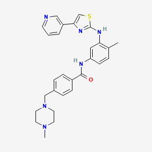
Masitinib

Masitinib; 790299-79-5; Masivet; AB1010; AB-1010;

CLASS:Immunomodulator
TARGET:KIT (a stem cell factor, also called c-KIT) receptor as well as select other tyrosine kinases
STATUS FOR MS:Phase III
COMMERCIAL:Under development by AB Science..Ab Science
4-((4-Methylpiperazin-1-yl)methyl)-N-(4-methyl-3-((4-(pyridin-3-yl)-1,3-thiazol-2-yl)amino)phenyl)benzamide
AB 1010
UNII-M59NC4E26P

4-((4-Methylpiperazin-1-yl)methyl)-N-(4-methyl-3-((4-(pyridin-3-yl)-1,3-thiazol-2-yl)amino)phenyl)benzamide

Regulatory and Commercial Status

STATUS FOR MS:Phase III
HIGHEST STATUS ACHIEVED (FOR ANY CONDITION):
Marketing Authorization Application for the treatment of pancreatic cancer has been filed with the European Medicines Agency (16 October 2012)
Marketing Authorization Application for the conditional approval in the treatment of pancreatic cancer has been accepted by the European Medicines Agency (30 October 2012)

Masitinib.png

Masitinib is a tyrosine-kinase inhibitor used in the treatment of mast cell tumors in animals, specifically dogs.[1][2] Since its introduction in November 2008 it has been distributed under the commercial name Masivet. It has been available in Europe since the second part of 2009. In the USA it is distributed under the name Kinavet and has been available for veterinaries since 2011.

Masitinib is being studied for several human conditions including cancers. It is used in Europe to fight orphan diseases.[3]

Mechanism of action

Masitinib inhibits the receptor tyrosine kinase c-Kit which is displayed by various types of tumour.[2] It also inhibits the platelet derived growth factor receptor (PDGFR) and fibroblast growth factor receptor (FGFR).

……………………..

http://www.google.com/patents/US7423055

Compound Synthesis

General: All chemicals used were commercial reagent grade products. Dimethylformamide (DMF), methanol (MeOH) were of anhydrous commercial grade and were used without further purification. Dichloromethane and tetrahydrofuran (THF) were freshly distilled under a stream of argon before use. The progress of the reactions was monitored by thin layer chromatography using precoated silica gel 60F 254, Fluka TLC plates, which were visualized under UV light. Multiplicities in 1H NMR spectra are indicated as singlet (s), broad singlet (br s), doublet (d), triplet (t), quadruplet (q), and multiplet (m) and the NMR spectrum were realized on a 300 MHz Bruker spectrometer.

3-Bromoacetyl-pyridine, HBr Salt

Dibromine (17.2 g, 108 mmol) was added dropwise to a cold (0° C.) solution of 3-acetyl-pyridine (12 g, 99 mmol) in acetic acid containing 33% of HBr (165 mL) under vigourous stirring. The vigorously stirred mixture was warmed to 40° C. for 2 h and then to 75° C. After 2 h at 75° C., the mixture was cooled and diluted with ether (400 mL) to precipitate the product, which was recovered by filtration and washed with ether and acetone to give white crystals (100%). This material may be recrystallised from methanol and ether.

IR (neat): 3108, 2047, 2982, 2559, 1709, 1603, 1221, 1035, 798 cm−1−1H NMR (DMSO-d6) δ=5.09 (s, 2H, CH2Br); 7.88 (m, 1H, pyridyl-H); 8.63 (m, 1H, pyridyl-H); 8.96 (m, 1H, pyridyl-H); 9.29 (m, 1H, pyridyl-H).

Methyl-[4-(1-N-methyl-piperazino)-methyl]-benzoate

To methyl-4-formyl benzoate (4.92 g, 30 mmol) and N-methyl-piperazine (3.6 mL, 32 mmol) in acetonitrile (100 mL) was added dropwise 2.5 mL of trifluoroacetic acid. The reaction mixture was stirred at room temperature for 1 h. After slow addition of sodium cyanoborohydride (2 g, 32 mmol), the solution was left stirring overnight at room temperature. Water (10 mL) was then added to the mixture, which was further acidified with 1N HCl to pH=6-7. The acetonitrile was removed under reduced pressure and the residual aqueous solution was extracted with diethyl ether (4×30 mL). These extracts were discarded. The aqueous phase was then basified (pH>12) by addition of 2.5N aqueous sodium hydroxyde solution. The crude product was extracted with ethyl acetate (4×30 mL). The combined organic layers were dried over MgSO4 and concentrated under reduced pressure to afford a slightly yellow oil which became colorless after purification by Kugelrohr distillation (190° C.) in 68% yield.

IR(neat): 3322, 2944, 2802, 1721, 1612, 1457, 1281, 1122, 1012—1H NMR(CDCl3) δ=2.27 (s, 3H, NCH3); 2.44 (m, 8H, 2×NCH2CH2N); 3.53 (s, 2H, ArCH2N); 3.88 (s, 3H, OCH3); 7.40 (d, 2H, J=8.3 Hz, 2×ArH); 7.91 (d, 2H, J=8.3 Hz, 2×ArH)—3C NMR (CDCl3) δ=45.8 (NCH3); 51.8 (OCH3); 52.9 (2×CH2N); 54.9 (2×CH2N); 62.4 (ArCH2N); 128.7 (2×ArC); 129.3 (2×ArC); 143.7 (ArC); 166.7 (ArCO2CH3)-MS CI (m/z) (%) 249 (M+1, 100%).

2-Methyl-5-tert-butoxycarbonylamino-aniline

A solution of di-tert-butyldicarbonate (70 g, 320 mmol) in methanol (200 mL) was added over 2 h to a cold (−10° C.) solution of 2,4-diaminotoluene (30 g, 245 mmol) and triethylamine (30 mL) in methanol (15 mL). The reaction was followed by thin layer chromatography (hexane/ethyl acetate, 3:1) and stopped after 4 h by adding 50 mL of water. The mixture was concentrated in vacuo and the residue was dissolved in 500 mL of ethyl acetate. This organic phase was washed with water (1×150 mL) and brine (2×150 mL), dried over MgSO4, and concentrated under reduced pressure. The resulting light brown solid was washed with small amounts of diethyl ether to give off-white crystals of 2-methyl-5-tert-butoxycarbonylamino-aniline in 67% yield.

IR (neat): 3359; 3246; 2970; 1719; 1609; 1557; 1173; 1050 cm−11H NMR (CDCl3): δ=1.50 (s, 9H, tBu); 2.10 (s, 3H, ArCH3); 3.61 (br s, 2H, NH2); 6.36 (br s, 1H, NH); 6.51 (dd, 1H, J=7.9 Hz, 2.3 Hz, ArH); 6.92 (d, 1H, J=7.9 Hz, ArH); 6.95 (s, 1H, ArH)—13C NMR (CDCl3) δ=16.6 (ArCH3); 28.3 (C(CH3)3); 80.0 (C(CH3)3); 105.2 (ArC); 108.6 (ArC); 116.9 (ArC); 130.4 (ArC—CH3); 137.2 (ArC—NH); 145.0 (ArC—NH2); 152.8 (COOtBu) MS ESI (m/z) (%): 223 (M+1), 167 (55, 100%).

N-(2-methyl-5-tert-butoxycarbonylamino)phenyl-thiourea

Benzoyl chloride (5.64 g, 80 mmol) was added dropwise to a well-stirred solution of ammonium thiocyanate (3.54 g, 88 mmol) in acetone (50 mL). The mixture was refluxed for 15 min, then, the hydrobromide salt of 2-methyl-5-tert-butoxycarbonylamino-aniline (8.4 g, 80 mmol) was added slowly portionswise. After 1 h, the reaction mixture was poured into ice-water (350 mL) and the bright yellow precipitate was isolated by filtration. This crude solid was then refluxed for 45 min in 70 mL of 2.5 N sodium hydroxide solution. The mixture was cooled down and basified with ammonium hydroxide. The precipitate of crude thiourea was recovered by filtration and dissolved in 150 mL of ethyl acetate. The organic phase was washed with brine, dried over Na2SO4, and concentrated under reduced pressure. The residue was purified by column chromatography (hexane/ethyl acetate, 1:1) to afford 63% of N-(2-methyl-5-tert-butoxycarbonylamino)phenyl-thiourea as a white solid.

IR (neat): 3437, 3292, 3175, 2983, 1724, 1616, 1522, 1161, 1053 cm−1— 1H NMR (DMSO-d6) δ=1.46 (s, 9H, tBu); 2.10 (s, 3H, ArCH3); 3.60 (br s, 2H, NH2); 7.10 (d, 1H, J=8.29 Hz, ArH); 7.25 (d, 1H, J=2.23 Hz, ArH); 7.28 (d, 1H, J=2.63 Hz, ArH); 9.20 (s, 1H, ArNH); 9.31 (s, 1H, ArNH)—13C NMR (DMSO-d6) δ=25.1 (ArCH3); 28.1 (C(CH3)3); 78.9 (C(CH3)3); 16.6 (ArC); 117.5 (ArC); 128.0 (ArC); 130.4 (ArC—CH3); 136.5 (ArC—NH); 137.9 (ArC—NH); 152.7 (COOtBu); 181.4 (C═S)—MS CI(m/z): 282 (M+1, 100%); 248 (33); 226 (55); 182 (99); 148 (133); 93 (188).

2-(2-methyl-5-tert-butoxycarbonylamino)phenyl-4-(3-pyridyl)-thiazole

A mixture of 3-bromoacetyl-pyridine, HBr salt (0.81 g, 2.85 mmol), N-(2-methyl-5-tert-butoxycarbonylamino)phenyl-thiourea (0.8 g, 2.85 mmol) and KHCO3 (˜0.4 g) in ethanol (40 mL) was heated at 75° C. for 20 h. The mixture was cooled, filtered (removal of KHCO3) and evaporated under reduced pressure. The residue was dissolved in CHCl3 (40 mL) and washed with saturated aqueous sodium hydrogen carbonate solution and with water. The organic layer was dried over Na2SO4 and concentrated. Colum chromatographic purification of the residue (hexane/ethyl acetate, 1:1) gave the desired thiazole in 70% yield as an orange solid

IR(neat): 3380, 2985, 2942, 1748, 1447, 1374, 1239, 1047, 938—1H NMR (CDCl3) δ=1.53 (s, 9H, tBu); 2.28 (s, 3H, ArCH3); 6.65 (s, 1H, thiazole-H); 6.89 (s, 1H); 6.99 (dd, 1H, J=8.3 Hz, 2.3 Hz); 7.12 (d, 2H, J=8.3 Hz); 7.35 (dd, 1H, J=2.6 Hz, 4.9 Hz); 8.03 (s, 1H); 8.19 (dt, 1H, J=1.9 Hz, 7.9 Hz); 8.54 (br s, 1H, NH); 9.09 (s, 1H, NH)—13C NMR (CDCl3) δ=18.02 (ArCH3); 29.2 (C(CH3)3); 81.3 (C(CH3)3); 104.2 (thiazole-C); 111.6; 115.2; 123.9; 124.3; 131.4; 132.1; 134.4; 139.5; 148.2; 149.1; 149.3; 153.6; 167.3 (C═O)—MS Cl (m/z) (%): 383 (M+1, 100%); 339 (43); 327 (55); 309 (73); 283 (99); 71 (311).

2-(2-methyl-5-amino)phenyl-4-(3-pyridyl)-thiazole

2-(2-methyl-5-tert-butoxycarbonylamino)phenyl-4-(3-pyridyl)-thiazole (0.40 g, 1.2 mmol) was dissolved in 10 mL of 20% TFA/CH2Cl2. The solution was stirred at rool temperature for 2 h, then it was evaporated under reduced pressure. The residue was dissolved in ethyl acetate. The organic layer was washed with aqueous 1N sodium hydroxide solution, dried over MgSO4, and concentrated to afford 2-(2-methyl-5-amino)phenyl-4-(3-pyridyl)-thiazole as a yellow-orange solid in 95% yield. This crude product was used directly in the next step.

A 2M solution of trimethyl aluminium in toluene (2.75 mL) was added dropwise to a cold (0° C.) solution of 2-(2-methyl-5-amino)phenyl-4-(3-pyridyl)-thiazole (0.42 g, 1.5 mmol) in anhydrous dichloromethane (10 mL) under argon atmosphere. The mixture was warmed to room temperature and stirred at room temperature for 30 min. A solution of methyl-4-(1-N-methyl-piperazino)-methyl benzoate (0.45 g, 1.8 mmol) in anhydrous dichloromethane (1 mL) and added slowly, and the resulting mixture was heated at reflux for 5 h. The mixture was cooled to 0° C. and quenched by dropwise addition of a 4N aqueous sodium hydroxide solution (3 mL). The mixture was extracted with dichloromethane (3×20 mL). The combined organic layers were washed with brine (3×20 mL) and dried over anhydrous MgSO4. (2-(2-methyl-5-amino)phenyl-4-(3-pyridyl)-thiazole) is obtained in 72% after purification by column chromatography (dichloromethane/methanol, 3:1)

IR (neat): 3318, 2926, 1647, 1610, 1535, 1492, 1282, 1207, 1160, 1011, 843—

1H NMR (CDCl3) δ=2.31 (br s, 6H, ArCH3+NCH3); 2.50 (br s, 8H, 2×NCH2CH2N); 3.56 (s, 2H, ArCH2N); 6.89 (s, 1H, thiazoleH); 7.21-7.38 (m, 4H); 7.45 (m, 2H); 7.85 (d, 2H, J=8.3 Hz); 8.03 (s, 1H); 8.13 (s, 1H); 8.27 (s, 1H); 8.52 (br s, 1H); 9.09 (s, 1H, NH)—

13C NMR (CDCl3) δ 17.8 (ArCH3); 46.2 (NCH3); 53.3 (NCH2); 55.3 (NCH2); 62.8 (ArCH2N); 99.9 (thiazole-C); 112.5; 123.9; 125.2; 127.5; 129.6; 131.6; 133.7; 134.0; 137.6; 139.3; 142.9; 148.8; 149.1; 166.2 (C═O); 166.7 (thiazoleC-NH)—

MS CI (m/z) (%): 499 (M+H, 100%); 455 (43); 430 (68); 401 (97); 374 (124); 309 (189); 283 (215); 235 (263); 121 (377); 99 (399).

………………………


http://www.google.com/patents/WO2012136732A1?cl=en

In a preferred embodiment of the above-depicted treatment, the active ingredient masitinib is administered in the form of masitinib mesilate; which is the orally bioavailable mesylate salt of masitinib – CAS 1048007-93-7 (MsOH); C28H30N6OS.CH3SO3H; MW 594.76:

Figure imgf000031_0001


http://www.google.com/patents/WO2004014903A1?cl=en

Figure imgf000021_0001

003 : 4-(4-Methyl-piperazin-l-ylmethyl)-N-[3-(4-pyridin-3-yl-thiazol-2-ylamino)- phenyl] -benzamide

4-(4-Methyl-piperazin-l-yl)-N-[4-methyl-3-(4-pyridin-3-yl-thiazol-2-ylmethyl)- phenyl] -benzamide

Figure imgf000053_0001

beige brown powder mp : 128-130°C

1H RMN (DMSO-d6) δ = 2.15 (s, 3H) ; 2.18 (s, 3H) ; 2.35-2.41 (m, 4H) ; 3.18-3.3.24 (m, 4H) ; 6.94 (d, J = 8.9 Hz, 2H) ; 7.09 (d, J = 8.4 Hz, IH) ; 7.28-7.38 (m, 3H) ; 7.81 (d, J = 8.9 Hz, 2H) ; 8.20-8.25 (m, IH) ; 8.40 (dd, J = 1.6 Hz, J = 4.7 , IH) ; 8.48 (d, J = 1.9 Hz, IH) ; 9.07 (d, J = 1.5 Hz, IH) ; 9.35 (s, IH) ; 9.84 (s, IH)

……………

http://www.google.com/patents/WO2008098949A2?cl=en

EXAMPLE 4 N- [4-Methyl-3 -(4-pyridin-3 -yl-thiazol-2-ylamino)-phenyl] -benzamide derivatives

Method A In a reactor and under low nitrogen pressure, add 4-Methyl-N3-(4-pyridin-3-yl-thiazol- 2-yl)-benzene-l,3-diamine (95 g, 336.45 mmol), dichloromethane (2 L). To this suspension cooled to temperature of 5°C was added dropwise 2M/n-hexane solution of trimethylaluminium (588 mL). The reaction mixture was brought progressively to 15°C, and maintained for 2 h under stirring. 4-(4-Methyl-piperazin-l-ylmethyl)-benzoic acid methyl ester (100 g, 402.71 mmol) in dichloromethane (200 mL) was added for 10 minutes. After 1 h stirring at room temperature, the reaction mixture was heated to reflux for 20 h and cooled to room temperature. This solution was transferred dropwise via a cannula to a reactor containing 2N NaOH (2.1 L) cooled to 5°C. After stirring for 3 h at room temperature, the precipitate was filtered through Celite. The solution was extracted with dichloromethane and the organic layer was washed with water and saturated sodium chloride solution, dried over MgSO4 and concentrated under vacuum. The brown solid obtained was recrystallized from /-Pr2O to give 130.7 g (78%) of a beige powder.

Method B Preparation of the acid chloride

To a mixture of 4-(4-Methyl-piperazin-l-ylmethyl)-benzoic acid dihydrochloride (1.0 eq), dichloromethane (7 vol) and triethylamine (2.15 eq), thionyl chloride (1.2 eq) was added at 18-28°C . The reaction mixture was stirred at 28-32°C for 1 hour. Coupling of acid chloride with amino thiazole To a chilled (0-50C) suspension of 4-Methyl-N3-(4-pyridin-3-yl-thiazol-2-yl)-benzene- 1,3-diamine (0.8 eq) and thiethylamine (2.2 eq) in dichloromethane (3 vol), the acid chloride solution (prepared above) was maintaining the temperature below 5°C. The reaction mixture was warmed to 25-300C and stirred at the same temperature for 1O h. Methanol (2 vol) and water (5 vol) were added to the reaction mixture and stirred. After separating the layers, methanol (2 vol), dihloromethane (5 vol) and sodium hydroxide solution (aqueous, 10%, till pH was 9.5-10.0) were added to the aqueous layer and stirred for 10 minutes. The layers were separated. The organic layer was a washed with water and saturated sodium chloride solution. The organic layer was concentrated and ethanol (2 vol) was added and stirred. The mixture was concentrated. Ethanol was added to the residue and stirred. The product was filtered and dried at 50-550C in a vaccum tray drier. Yield = 65-75%.

Method C

To a solution of 4-methyl-N3-(4-pyridin-3-yl-thiazol-2-yl)-benzene-l,3-diamine (1.0 eq) in DMF (20 vol) were added successively triethylamine (5 eq), 2-chloro-l- methylpyridinium iodide (2 eq) and 4-(4-methyl-piperazin-l-ylmethyl)-benzoic acid (2 eq). The reaction mixture was stirred for 7 h at room temperature. Then, the mixture was diluted in diethyl ether and washed with water and saturated aqueous NaHCO3, dried over Na2SO4 and concentrated. The crude product was purified by column chromatography using an elution of 100% EtOAc to give a yellow solid.

Yield = 51%.

1H NMR (CDCl3) : δ = 9.09 (IH, s, NH); 8.52 (IH, br s); 8.27 (IH, s); 8.13 (IH, s);

8.03 (IH, s); 7.85 (2H, d, J= 8.3Hz); 7.45 (2H, m); 7.21-7.38 (4H, m); 6.89 (IH, s);

3.56 (2H, s); 2.50 (8H, br s); 2.31 (6H, br s).

MS (CI) m/z = 499 (M+H)+.

An additional aspect of the present invention relates to a particular polymorph of the methanesulfonic acid salt of N-[4-Methyl-3-(4-pyridin-3-yl-thiazol-2-ylamino)-phenyl]- benzamide of formula (IX).

Figure imgf000023_0001

(VI)

Hereinafter is described the polymorph form of (IX) which has the most advantageous properties concerning processability, storage and formulation. For example, this form remains, dry at 80% relative humidity and thermodynamically stable at temperatures below 2000C.

The polymorph of this form is characterized by an X-ray diffraction pattern illustrated in FIG.I, comprising characteristic peaks approximately 7.269, 9.120, 11.038, 13.704, 14.481, 15.483, 15.870, 16.718, 17.087, 17.473, 18.224, 19.248, 19.441, 19.940, 20.441, 21.469, 21.750, 22.111, 23.319, 23.763, 24.120, 24.681, 25.754, 26.777, 28.975, 29.609, 30.073 degrees θ, and is also characterized by differential scanning calorimetry (DSC) illustrated in FIG.II, which exhibit a single maximum value at approximately 237.49 ± 0.3 0C. X-ray diffraction pattern is measured using a Bruker AXS (D8 advance). Differential scanning calorimetry (DSC) is measured using a Perking Elmer Precisely (Diamond DSC).

This polymorph form can be obtained by treatement of 4-(4-Methyl-piperazin-l- ylmethyl)-N-[4-methyl-3-(4-pyridin-3-yl-thiazol-2-ylamino)-phenyl]-benzamide with 1.0 to 1.2 equivalent of methanesulfonic acid, at a suitable temperature, preferably between 20-800C.

The reaction is performed in a suitable solvent especially polar solvent such as methanol or ethanol, or ketone such as acetone, or ether such as diethylether or dioxane, or a mixture therof. This invention is explained in example given below which is provided by way of illustration only and therefore should not be construed to limit the scope of the invention. Preparation of the above-mentioned polymorph form of 4-(4-Methyl-piperazin-l- ylmethyl)-N- [4-methyl-3 -(4-pyridin-3 -yl-thiazol-2-ylamino)-phenyl] -benzamide methanesulfonate .

4-(4-Methyl-piperazin- 1 -ylmethyl)-N- [4-methyl-3 -(4-pyridin-3 -yl-thiazol-2-ylamino) phenyl] -benzamide (1.0 eq) was dissolved in ethanol (4.5 vol) at 65-700C. Methanesulfonic acid (1.0 eq) was added slowly at the same temperature. The mixture was cooled to 25-300C and maintained for 6 h. The product was filtered and dried in a vacuum tray drier at 55-600C. Yield = 85-90%. Starting melting point Smp = 236°C.


NMR PREDICT

CAS NO. 1048007-93-7, methanesulfonic acid,4-[(4-methylpiperazin-1-yl)methyl]-N-[4-methyl-3-[(4-pyridin-3-yl-1,3-thiazol-2-yl)amino]phenyl]benzamide H-NMR spectral analysis

methanesulfonic acid,4-[(4-methylpiperazin-1-yl)methyl]-N-[4-methyl-3-[(4-pyridin-3-yl-1,3-thiazol-2-yl)amino]phenyl]benzamide NMR spectra analysis, Chemical CAS NO. 1048007-93-7 NMR spectral analysis, methanesulfonic acid,4-[(4-methylpiperazin-1-yl)methyl]-N-[4-methyl-3-[(4-pyridin-3-yl-1,3-thiazol-2-yl)amino]phenyl]benzamide H-NMR spectrum

methanesulfonic acid,4-[(4-methylpiperazin-1-yl)methyl]-N-[4-methyl-3-[(4-pyridin-3-yl-1,3-thiazol-2-yl)amino]phenyl]benzamide NMR spectra analysis, Chemical CAS NO. 1048007-93-7 NMR spectral analysis, methanesulfonic acid,4-[(4-methylpiperazin-1-yl)methyl]-N-[4-methyl-3-[(4-pyridin-3-yl-1,3-thiazol-2-yl)amino]phenyl]benzamide C-NMR spectrum

CAS NO. 1048007-93-7, methanesulfonic acid,

4-[(4-methylpiperazin-1-yl)methyl]-N-[4-methyl-3-[(4-pyridin-3-yl-1,3-thiazol-2-yl)amino]phenyl]benzamide C-NMR spectral analysisPREDICT

References

  1. Hahn, K.A.; Oglivie, G.; Rusk, T.; Devauchelle, P.; Leblanc, A.; Legendre, A.; Powers, B.; Leventhal, P.S.; Kinet, J.-P.; Palmerini, F.; Dubreuil, P.; Moussy, A.; Hermine, O. (2008). “Masitinib is Safe and Effective for the Treatment of Canine Mast Cell Tumors”. Journal of Veterinary Internal Medicine 22 (6): 1301–1309. doi:10.1111/j.1939-1676.2008.0190.x. ISSN 0891-6640.
  2. Information about Masivet at the European pharmacy agency website
  3. Orphan designation for Masitinib at the European pharmacy agency website
WO2004014903A1 Jul 31, 2003 Feb 19, 2004 Ab Science 2-(3-aminoaryl)amino-4-aryl-thiazoles and their use as c-kit inhibitors
WO2008098949A2 Feb 13, 2008 Aug 21, 2008 Ab Science Process for the synthesis of 2-aminothiazole compounds as kinase inhibitors
EP1525200B1 Jul 31, 2003 Oct 10, 2007 AB Science 2-(3-aminoaryl)amino-4-aryl-thiazoles and their use as c-kit inhibitors
US7423055 Aug 1, 2003 Sep 9, 2008 Ab Science 2-(3-Aminoaryl)amino-4-aryl-thiazoles for the treatment of diseases
US20080207572 * Jul 13, 2006 Aug 28, 2008 Ab Science Use of Dual C-Kit/Fgfr3 Inhibitors for Treating Multiple Myeloma
Masitinib.svg
Systematic (IUPAC) name
4-[(4-Methylpiperazin-1-yl)methyl]-N-(4-methyl-3-{[4-(pyridin-3-yl)-1,3-thiazol-2-yl]amino}phenyl)benzamide
Clinical data
Trade names Masivet, Kinavet
AHFS/Drugs.com International Drug Names
Identifiers
790299-79-5
L01XE22
PubChem CID 10074640
ChemSpider 8250179
ChEMBL CHEMBL1908391
Chemical data
Formula C28H30N6OS
498.64 g/mol
Patent Submitted Granted
2-(3-Aminoaryl)amino-4-aryl-thiazoles for the treatment of diseases [US7423055] 2004-06-10 2008-09-09
2-(3-aminoaryl)amino-4-aryl-thiazoles and their use as c-kit inhibitors [US2005239852] 2005-10-27
Use of C-Kit Inhibitors for Treating Fibrosis [US2007225293] 2007-09-27
Use of Mast Cells Inhibitors for Treating Patients Exposed to Chemical or Biological Weapons [US2007249628] 2007-10-25
Use of c-kit inhibitors for treating type II diabetes [US2007032521] 2007-02-08
Use of tyrosine kinase inhibitors for treating cerebral ischemia [US2007191267] 2007-08-16
Use of C-Kit Inhibitors for Treating Plasmodium Related Diseases [US2008004279] 2008-01-03
Tailored Treatment Suitable for Different Forms of Mastocytosis [US2008025916] 2008-01-31
2-(3-AMINOARYL) AMINO-4-ARYL-THIAZOLES AND THEIR USE AS C-KIT INHIBITORS [US2008255141] 2008-10-16
Use Of C-Kit Inhibitors For Treating Inflammatory Muscle Disorders Including Myositis And Muscular Dystrophy [US2008146585] 2008-06-19
Patent Submitted Granted
Aminothiazole compounds as kinase inhibitors and methods of using the same [US8940894] 2013-05-10 2015-01-27
Aminothiazole compounds as kinase inhibitors and methods of using the same [US8492545] 2012-03-08 2013-07-23
Patent Submitted Granted
Use of Dual C-Kit/Fgfr3 Inhibitors for Treating Multiple Myeloma [US2008207572] 2008-08-28
PROCESS FOR THE SYNTHESIS OF 2-AMINOTHIAZOLE COMPOUNDS AS KINASE INHIBITORS [US8153792] 2010-05-13 2012-04-10
COMBINATION TREATMENT OF SOLID CANCERS WITH ANTIMETABOLITES AND TYROSINE KINASE INHIBITORS [US8227470] 2010-04-15 2012-07-24
Anti-IGF antibodies [US8580254] 2008-06-19 2013-11-12
COMBINATIONS FOR THE TREATMENT OF B-CELL PROLIFERATIVE DISORDERS [US2009047243] 2008-07-17 2009-02-19
TREATMENTS OF B-CELL PROLIFERATIVE DISORDERS [US2009053168] 2008-07-17 2009-02-26
Anti-IGF antibodies [US8318159] 2009-12-11 2012-11-27
SURFACE TOPOGRAPHIES FOR NON-TOXIC BIOADHESION CONTROL [US2010226943] 2009-08-31 2010-09-09
EGFR/NEDD9/TGF-BETA INTERACTOME AND METHODS OF USE THEREOF FOR THE IDENTIFICATION OF AGENTS HAVING EFFICACY IN THE TREATMENT OF HYPERPROLIFERATIVE DISORDERS [US2010239656] 2010-05-10 2010-09-23
ANTI CD37 ANTIBODIES [US2010189722] 2008-08-08 2010-07-29
United States National Library of Medicine

Note: Compound name must be entered under “Substance Identification” and then “Names and Synonyms” selected to view synonyms.

Kocic I, Kowianski P, Rusiecka I, Lietzau G, Mansfield C, Moussy A, Hermine O, Dubreuil P
Naunyn Schmiedebergs Arch Pharmacol. 2014 Oct 26. Epub 2014 Oct 26. PMID: 25344204.Abstract
AB SCIENCE HEADQUARTER
3, Avenue George V
75008 PARIS – FRANCE
Tel. : +33 (0)1 47 20 00 14
Fax. : +33 (0)1 47 20 24 11

P.S. : The views expressed are my personal and in no-way suggest the views of the professional body or the company that I represent.

P.S. : The views expressed are my personal and in no-way suggest the views of the professional body or the company that I represent.

P.S. : The views expressed are my personal and in no-way suggest the views of the professional body or the company that I represent.

TAJIKISTAN

Tajikistan – Wikipedia, the free encyclopedia

en.wikipedia.org/wiki/Tajikistan

The territory that now constitutes Tajikistan was previously home to several ancient cultures, including the city of Sarazm of the Neolithic and the Bronze Age, …

Map of tajikistan country.
The nature of Tajikistan. Nurek
Tajikistan. Pamiro-Alay.Zeravshan mountain range. Guzn village. Local people
Dushanbe, Tajikistan
Women carry water canisters near Gargara village, 110km south of Tajikistan’s capital, Dushanbe
Ancient Buddhist ruins, Ajina Teppa, Tajikistan
///////////

Share this:

Share

Treating the flu?

 Uncategorized  Comments Off on Treating the flu?
May 092015
 

Treating the flu?

They walked out together into the fine fall day, scuffling bright ragged leaves under their feet, turning their faces up to a generous sky really blue and spotless. At the first corner they waited for a funeral to pass, the mourners seated straight and firm as if proud in their sorrow. […] “It seems to be a plague,” said Miranda, “something out of the Middle Ages. Did you ever see so many funerals, ever?”
— from “Pale Horse, Pale Rider” by Katherine Anne Porter (1939)

And I looked, and behold a pale horse: and his name that sat on him was Death, and Hell followed with him.
— Revelations 6.8 (King James Version)


In 1918-1919 between 50 and 100 million people worldwide died from the flu. The “Spanish Flu” spread to nearly every part of the world with amazing speed, helped perhaps by the thousands of soldiers returning from Europe after the end of World War I. There was little that could be done to help the sick, and often people who were healthy one day were dead the next. The Spanish Flu was remarkable at the time in that it primarily killed young healthy adults, whereas most often it is very young children and elderly people who die from infectious disease.

Oddly enough, after going through two successive waves of infection and mortality, the Spanish Flu pandemic disappeared almost abruptly. By the end of the 20th century, it was almost forgotten, and influenza had come to be regarded as one of the many childhood diseases that most people went through without much difficulty.

The situation today is quite different. Everyone is now highly sensitized to the threat of influenza. Stories about the so-called “Bird Flu” and now the “Swine Flu” have appeared regularly on television and in newspapers. Our society is more mobile than ever before, and we have seen examples of the rapid spread of diseases worldwide in recent years. Population is far more dense than it was in 1918, and diseases spread and mutate in crowded cities around the world far faster than ever before. People are deeply concerned about the possibility of a new influenza pandemic that could rival the Spanish Flu.

On the other hand, we also now know much more about how to prevent and how to treat illnesses like influenza. The best way to slow or stop the spread of influenza is through public health measures – simple things like frequent hand washing and avoiding contact with infected people. In addition, immunization is an important protective measure if a safe and effective vaccine can be developed.

But what about treating people who are already infected? Because influenza is a viral disease, antibiotics that can deal with bacterial infections will not work. The story of how drugs to treat serious cases of influenza were developed shows how structural biology, biochemistry and synthetic organic chemistry work hand-in-hand to produce new and useful chemical substances. It remains to be seen if they can help in the event of a pandemic outbreak, which many people think is a question of “when” rather than “if”.


Treating the flu? Part 1: The Influenza Virus

Influenza is caused by RNA viruses of the family Orthomyxoviridae. These virions are roughly 80-120 microns in diameter. Their surfaces consist of a lipid bilayer derived from the membrane of the host cell, which is decorated by glycoproteins that project like spikes from the viral particle. About 80% of these spikes are hemagglutinin, a protein that facilitates binding the virion to a host cell. The remainder areneuraminidase, which is an enzyme that cleaves glycosidic linkages to the sugar neuraminic acid (also calledsialic acid).

You have probably heard the different strains of the flu virus (“serotypes”) referred to as “H1N1” or “H5N1”. These names refer to the different subtypes of the two surface glycoproteins, differences that distinguish the serotypes immunogenically.

There are several outstanding web sites that will tell you much more about the influenza virus. There is no point in just repeating what they contain here, so if you want more information you can follow the links below. Otherwise, click here to move to the next part of the drug development story.


Treating the flu? Part 2: Targets for therapy

A drug must act by binding to and modulating the activity of some target receptor or enzyme. Viruses do not present very many potential targets because they typically have only a few unique proteins coded in their genomes. Recall that viruses hi-jack the enzymes of the host cell to manufacture new virions.

The Influenza A genome consists of 8 strands of RNA:

1. The HA gene. It encodes the hemagglutinin.
2. The NA gene. It encodes the neuraminidase.
3. The NP gene encodes the nucleoprotein. Influenza A, B, and C viruses have different nucleoproteins.
4. The M gene encodes two proteins (using different reading frames of the RNA): a matrix protein M1 and an ion channel M2 spanning the lipid bilayer.
5. The NS gene encodes two different non-structural proteins that are found in the cytoplasm of the infected cell but not within the virion itself.
6. – 8. one RNA molecule (PA, PB1, PB2) for each of the 3 subunits of the RNA polymerase.

Drugs against Influenza A could potentially be developed to inhibit the activity of any of the products of the influenza genome, but in fact only drugs acting against the NA (neuraminidase) and the M2 (ion channel) proteins have been successfully developed to date.

The M2 inhibitors amantadine and rimantadine were the first effective drugs against influenza, but the M2 protein seems quite easy for the virus to modify so resistance rapidly develops against these drugs. The latest H1N1 virus that is causing pandemic concern is resistant to both amantadine and rimantadine. The drugs that are being used against current pandemic threat strains target the viral neuraminidase, and it is these that form the basis of our discussion on drug development.


Treating the flu? Part 3: Neuraminidase

This is only a very short description of this important enzyme. It assumes that you have some basic knowledge of what enzymes are and what they do. If you need more background information, your Biochemistry textbook or the Wikipedia article on enzymes are good places to start.

Recall that the surface of the influenza virion is covered with spikes of hemagglutinin and neuraminidase. Hemagglutinin is a protein that binds tightly to the sugar portions of various cell-surface glycoproteins by recognizing and binding the sugarsialic acid, which is also called N-acetyl neuraminic acid. Sialic acid is found at the terminus of the carbohydrate portions of many cell-surface glycoproteins and plays a key role in cell-cell and cell-virus binding. The human ABO blood-group antigens are examples of sialylated oligosaccharides that play an important role in medical biochemistry.

Hemagglutinin permits the influenza virus to attach to a host cell during the initial infection, which in turn causes the viral RNA to enter the cell by endocytosis. This is a common mechanism for infection and we know that many viruses including HIV as well as parasites such as the Plasmodium that causes malaria attack host cells via their cell-surface carbohydrates. However, the tight grip of viral hemagglutinin on cell-surface sialic acid is a problem when new viral particles need to break away from the host cell.

The neuraminidase on the surface of the virion is necessary for new viral particles to break away from the host cell. Neuraminidase is a glycosidase (an enzyme that catalyzes the hydrolysis of glycosidic linkages) that specifically promotes the cleavage of sialic acid from glycoprotein saccharide chains. When the glycosidic linkage is cleaved by hydrolysis, the sialic acid falls off the cell surface. The viral particle is now no longer tethered to the host cell and can move off to infect other cells.

If the activity of neuraminidase is blocked, the new virions remain bound to the host cell and viral reproduction is prevented. You can view a Flash animation showing this concept here.


The chemical structure of sialic acid or N-acetyl neuraminic acid.


The structure of the influenza A neuraminidase N9 bound to an analogue of sialic acid has been determined by X-ray crystallography, and a simplified ribbon diagram is shown here. The amino acid chains are represented by the yellow ribbons, and the bound inhibitor as well as some key side chain groups are shown in ball-and-stick format. The broad arrows designate regions in which the amino acid chains form a “beta sheet” structure, with the arrow heads indicating the C-terminal end of the sheet. Cylindrical sections represent “random coil” regions of the amino acid sequence. Notice that there is essentially no helical structure in this enzyme. This image shows only one sub-unit of the biologically active form of the enzyme which is actually a tetramer of identical sub-units.

The binding site of the enzyme does not vary from strain to strain. It consists of 18 amino acid residues of which 12 are in direct contact with the bound sialic acid analogue (and presumably with sialic acid in catalytically active situations). Four of these 12 are positively-charged arginines, while another 4 are negatively-charged glutamic and aspartic acid residues. The remainder are neutral (tyrosine, asparagine, isoleucine and tryptophan).

If you visit the RCSB Protein Data Bank you can find X-ray structures of many neuraminidases – this one is indexed under the code “1nna“. The details of the structure are discussed in the original paper by Bossart-Whitaker et al. cited below.


A schematic diagram of the 3-D structure of neuraminidase showing how it binds to sialic acid.

Mark von Itzstein and coworkers (then at the Monash University Victorian College of Pharmacy in Melbourne Australia and now at the Institute for Glycomics at Australia’s Griffith University) studied the mechanism of sialic acid hydrolysis catalyzed by influenza A N9 neuraminidase. This enzyme is what is called a retaining glycosidase because if the starting glycoside has the α-configuration (as shown) then the product that is formed will also have the α-configuration. In common with many glycosidase enzymes, its active site features a pair of carboxyl residues (Asp 151 and Glu 277 in the N9 neuraminidase they studied) which play central roles in the enzyme’s catalytic mechanism. The proposed mechanism is shown below.

There are two important transition states shown in this mechanism, the first for the actual cleavage of the C-O bond leading to loss of the ROH fragment and the second for the formation of a new C-OH bond. In the first transition state, notice how the enzyme assists the ionization of a water molecule, the transfer of its proton to the leaving OR group, and stabilizes the transient positive charge on the ring oxygen.

With knowledge of how the enzyme functioned, von Itzstein decided that a compound that looked like the carbohydrate in that key first transition state would be a good candidate for an anti-influenza drug that would function by preventing the release of viral particles from infected cells. Click here to go to the next stage in the story – synthesizing and testing a new compound.


Wikipedia article about neuraminidase.

The story of how neuraminidase was identified as a target for anti-influenza drug development is briefly outlined by Graeme Laver, one of the key researchers in this field. You can read his March 2007 article in Education in Chemistry here.

Bossart-Whitaker, P.; Carson, M.; Babu, Y.S.; Smith, C.D.; Laver, W.G.; Air, G.M. J. Mol. Biol. 1993232, 1069–1083. (Link requires valid U of Manitoba Library ID).

von Itzstein, M. et al. Nature 1993363, 418-423. (Link requires valid U of Manitoba Library ID).


Treating the flu? Part 4: Developing Neuraminidase Inhibitors

Zanamivir (Relenza)

Note: this document should not be taken as any form of endorsement of the substances mentioned or as a recommendation for treatment.

With the information gained from structural and mechanistic studies on influenza A neuraminidase, von Itzstein and his team set out to devise and synthesize a stable molecule that looked sufficiently like the transition state to bind very tightly to the enzyme, thus inhibiting it. Recall that a transition state is not a stable isolable molecule, but it is possible to mimic the geometry of a proposed transition state with other chemical structures. These are called transition state analogues.The proposed transition state for hydrolysis of glycosidic bonds to sialic acid catalyzed by influenza A neuraminidase.

The proposed transition state for glycosidic bond cleavage in the mechanism previously outlined is shown here. Recall that for clarity the sugar structure has been simplified. It is evident that the reactive centre of the sugar ring is planar in this transition state. It is not possible to make a stable structure that has a double bond between position 2 and the ring oxygen similar to the partial double bond in the transition structure. Thus, von Itzstein et al. decided that a good inhibitor needed a double bond between positions 2 and 3 – that is, it should be a 2,3-dehydro derivative of sialic acid.

They also concluded that a strongly basic guanidino group should replace the hydroxyl at C-4 in the sialic acid structure. This would be positively charged at physiological pH and would bind strongly to a region of negative charge in the active site.

They synthesized and tested the structure shown in 1989 and found that it was indeed a potent and very selective inhibitor of influenza neuraminidase. Their synthetic route, published in the journal Carbohydrate Research in 1994, is shown below.

Although some of the reagents used in this synthesis may be unfamiliar, organic chemistry students should be able to recognize what is going on in each step. In the first step shown, the Lewis acid boron trifluoride etherate promotes an internal SN2 reaction in which the carbonyl of the acetamide displaces the acetate ester to form the new ring. Notice the inversion of configuration at C4. This is then subjected to another SN2 reaction in which the nucleophile is the azide anion N3. The reagent is trimethylsilyl azide, which also provides mildly Lewis acidic activation for the displacement. Azide groups are excellent precursors for amines, and the reduction of the azide is easily carried out. You can see that some care must be taken here, since if the reaction is left too long the hydrogenation of the alkene will also occur. Simple alkaline hydrolysis removes the methyl ester and the acetate ester protecting groups, and then the amino group is converted into the desired guanidino function using formamidine sulfonic acid. This provided the desired neuraminidase inhibitor 4-deoxy-4-guanidino-2,3-dehydro-N-acetyl neuraminic acid, which ultimately has become the anti-influenza drug zanamivir (sold under the trade name Relenza by GlaxoSmithKline).

You can see how well zanamivir fits into the active site of influenza A neuraminidase from the X-ray crystal structure obtained by Zu et al. and indexed in theProtein Data Bank as 3b7e. This is an interesting structure because the enzyme is the neuraminidase from the A/Brevig Mission/1/1918 H1N1 strain, one of the viruses that caused the 1918 Spanish Flu. The genome of this virus was obtained from the frozen body of a woman who died in the Alaskan village of Brevig Missionin 1918. An interesting New York Times article describes the discovery of this virus (and incidentally the Johan Hultin who found the virus is no relation to Dr. Hultin!). It is another variation of the H1N1 strain that is at the centre of the 2009/2010 concern about Swine Flu.

The ribbon diagram has simplified the enzyme structure considerably – only those amino acids near the active site are shown, and only the most important ones that interact with zanamivir have their sidechains drawn. The drug molecule is shown in a space-filling representation in which oxygen is red, nitrogen is blue and carbon is white. Hydrogens are not shown. The diagram places the carboxylate group of zanamivir at the 6 o’clock position, while the guanidinium group is projecting backwards deep into the binding site. The hydroxylated sidechain is projecting forward at about 9 o’clock. The schematic drawing (based on a diagram from the book by Levy and Fugedi referenced below) shows the key contacts between the enzyme and the drug.

Numerous other synthetic routes to zanamivir have been published since the original synthesis shown here, and you can be very sure that the industrial synthesis isquite different. The problem with Zanamivir is that it cannot be administered orally. Because the guanidino group is strongly basic, if it were taken orally it would be protonated in the stomach. The resulting positively-charged structure could not be taken up from the gut. Zanamivir is usually administered by inhalation, but this is not as acceptable to many people as a pill would be, and does not give a particularly high level of bioavailability.

Given this problem with zanamivir, it is not surprising that others tried to find similar compounds to inhibit influenza neuraminidase that could be orally administered. Click here to find out about the second-generation drug oseltamivir (Tamiflu).


von Itzstein, M.; Wu, W.-Y.; Jin, B. Carbohydrate Research 1994259, 301-305.

Taylor, N.R.; von Itzstein, M. J. Med. Chem. 199437, 616–624.

Magano, J. Chem. Rev. 2009, in press. (You must have a valid U of Manitoba library ID to access the full-text article)

Xu, X.; Zhu, X.; Dwek, R.A.; Stevens, J.; Wilson, I.A. J.Virol. 2008, 82, 10493-10501.

Levy, D.; Fugedi, P. (Eds.) The Organic Chemistry of Sugars, CRC/Taylor & Francis: 2006.


Treating the flu? Part 4: Developing Neuraminidase Inhibitors

Other neuraminidase inhibitors

Research and development of new anti-influenza drugs has not stopped. The need for more effective drugs remains a powerful incentive for academic and industrial scientists, and there is of course a strong profit motive as well.


One compound that is now in clinical trials is peramivir, under development by BioCryst Pharmaceuticals. If you look at the structure of peramivir, you can see its family resemblance to other neuraminidase inhibitors. However, peramivir must be administered by injection because it has rather poor oral bioavailability. In fact, peramivir was initially developed by Johnson and Johnson but was abandoned because it was not orally active. Renewed interest in it as an injectable drug may be because only the most severe cases of influenza really need antiviral therapy, and such patients are likely already hospitallized.

Another new compound is CS-8958, from Japan’s Daiichi Sankyo Co. Ltd. This compound is structurally very similar to zanamivir, differing only in the functionalization of the hydroxylated sidechain.

CS-8958 is a prodrug and not the active form. The octyl ester group is hydrolyzed in the liver, releasing the active neuraminidase inhibitor which only differs from zanamivir in having a methyl ether at the C7 position rather than a hydroxyl group. The main advantage of CS-8958 is that it is long-acting. Oseltamivir and zanamivir must be taken twice daily, but in a clinical study a single inhaled treatment with CS-8958 gave the same anti-influenza effect as twice-daily doses of oseltamivir over 5 days.

Share

Design and Synthesis of Pyridinylisoxazoles and Their Anticancer Activities

 Uncategorized  Comments Off on Design and Synthesis of Pyridinylisoxazoles and Their Anticancer Activities
Apr 102015
 
YANG Hongliang, XU Guoxing, BAO Meiying, ZHANG Dapeng, LI Zhiwei, PEI Yazhong
Design and Synthesis of Pyridinylisoxazoles and Their Anticancer Activities
2014 Vol. 35 (12): 2584-2592 [Abstract] ( 781 ) [HTML 0KB] [PDF 2464KB] (116 )
doi10.7503/cjcu20140333

Chemical Journal of Chinese Universities  2014Vol. 35  Issue (12): 2584-2592    DOI: 10.7503/cjcu20140333

Abstract  Based on the X-ray co-crystal structures of reported allosteric kinase inhibitors bound to their corresponding protein kinases, a pharmacophore model was proposed. To examine the validity of this hypothesis, 21 new pyridinylisoxazole derivatives were designed and synthesized. Their structures were confirmed using 1H NMR, 13C NMR and MS data. Their inhibitory effects against human breast cancer cell(MCF-7) proliferation were evaluated. Preliminary results indicated that some of these pyridinylisoxazole derivatives possess potent anti-proliferative activities, with IC50 data in the micromolar range. The mechanism-of-action of these compounds is under investigation.

Cite this article:
Design and Synthesis of Pyridinylisoxazoles and Their Anticancer Activities
YANG Hongliang1, XU Guoxing1, BAO Meiying2, ZHANG Dapeng1, LI Zhiwei1, PEI Yazhong1
1. The Center for Combinatorial Chemistry and Drug Discovery, School of Pharmaceutical Sciences, Jilin University, Changchun 130021, China;
2. Changchun Discovery Sciences Co. Ltd., Changchun 130012, China
YANG Hongliang,XU Guoxing,BAO Meiying et al. Design and Synthesis of Pyridinylisoxazoles and Their Anticancer Activities[J]. Chemical Journal of Chinese Universities, 2014, 35(12): 2584-2592.
URL:
http://www.cjcu.jlu.edu.cn/EN/10.7503/cjcu20140333     OR     http://www.cjcu.jlu.edu.cn/EN/Y2014/V35/I12/2584


Displaying image006.png


Displaying image007.png


Displaying image010.png


Share
Follow

Get every new post on this blog delivered to your Inbox.

Join other followers: