AUTHOR OF THIS BLOG

DR ANTHONY MELVIN CRASTO, WORLDDRUGTRACKER

Cipla to invest in South Africa’s first biosimilars production facility

 INDIA, MONOCLONAL ANTIBODIES  Comments Off on Cipla to invest in South Africa’s first biosimilars production facility
Jul 142016
 

Cipla to invest in South Africa’s first biosimilars production facility
Indian-based pharmaceutical and biotechnology company Cipla will invest more than R1.3bn ($19.34m) in the first advanced biotech manufacturing facility in South Africa for the production of biosimilars.

Indian-based pharmaceutical and biotechnology company Cipla will invest more than R1.3bn ($19.34m) in the first advanced biotech manufacturing facility in South Africa for the production of biosimilars.

The investment will be carried out by South African subsidiary Cipla BioTec…………………cont

read at

http://www.pharmaceutical-technology.com/news/newscipla-invest-south-africas-first-biosimilars-production-facility-4945516?WT.mc_id=DN_News

Cipla Managing director and global CEO Subhanu Saxena


Dr Y.K. Hamied,

Department of Trade and Industries Special Economic Zone of Dube Tradeport, DURBAN, SOUTHAFRICA


///Cipla, South Africa, biosimilars,  production facility, Dube Tradeport, Cipla BioTec Pvt Ltd, Durban, SOUTHAFRICA

Share

タバルマブ(遺伝子組換え) Tabalumab

 MONOCLONAL ANTIBODIES, Uncategorized  Comments Off on タバルマブ(遺伝子組換え) Tabalumab
Jun 272016
 

Tabalumab


タバルマブ(遺伝子組換え)
Tabalumab (Genetical Recombination)

[1143503-67-6]

Tabalumab (LY 2127399) is an anti-B-cell activating factor (BAFF) human monoclonal antibody designed for the treatment of autoimmune diseases and B cell malignancies.[1][2] Tabalumab was developed by Eli Lilly and Company.

A phase III clinical trial for rheumatoid arthritis was halted in Feb 2013.[3] In September 2014, a second phase III trial focussing on treating systemic lupus erythematosus, was terminated early as the study failed to meet its primary endpoint.[4]


References


abalumab
Monoclonal antibody
Type Whole antibody
Source Human
Target BAFF
Identifiers
CAS Number 1143503-67-6 
ATC code none
ChemSpider none
Chemical data
Formula C6518H10008N1724O2032S38
Molar mass 146.25 kg/mol

////////////タバルマブ ,  遺伝子組換え, Tabalumab, 1143503-67-6, antibody, Monoclonal antibody

Share

Perspectives on Anti-Glycan Antibodies Gleaned from Development of a Community Resource Database

 MONOCLONAL ANTIBODIES  Comments Off on Perspectives on Anti-Glycan Antibodies Gleaned from Development of a Community Resource Database
Jun 112016
 
Abstract Image

Antibodies are used extensively for a wide range of basic research and clinical applications. While an abundant and diverse collection of antibodies to protein antigens have been developed, good monoclonal antibodies to carbohydrates are much less common. Moreover, it can be difficult to determine if a particular antibody has the appropriate specificity, which antibody is best suited for a given application, and where to obtain that antibody. Herein, we provide an overview of the current state of the field, discuss challenges for selecting and using antiglycan antibodies, and summarize deficiencies in the existing repertoire of antiglycan antibodies. This perspective was enabled by collecting information from publications, databases, and commercial entities and assembling it into a single database, referred to as the Database of Anti-Glycan Reagents (DAGR). DAGR is a publicly available, comprehensive resource for anticarbohydrate antibodies, their applications, availability, and quality

Monoclonal antibodies have transformed biomedical research and clinical care. In basic research, these proteins are used widely for a myriad of applications, such as monitoring/detecting expression of biomolecules in tissue samples, activating or antagonizing various biological pathways, and purifying antigens. To illustrate the magnitude and importance of the antibody reagent market, one commercial supplier sells over 50 000 unique monoclonal antibody clones. In a clinical setting, antibodies are used frequently as therapeutic agents and for diagnostic applications. As a result, monoclonal antibodies are a multibillion dollar industry, with antibody therapeutics estimated at greater than $40 billion annually, diagnostics at roughly $8 billion annually, and antibody reagents at $2 billion annually as of 2012

Perspectives on Anti-Glycan Antibodies Gleaned from Development of a Community Resource Database

Chemical Biology Laboratory, Center for Cancer Research, National Cancer Institute, Frederick, Maryland 21702, United States
ACS Chem. Biol., Article ASAP
DOI: 10.1021/acschembio.6b00244
Publication Date (Web): May 25, 2016
Copyright © 2016 American Chemical Society

ACS Editors’ Choice – This is an open access article published under an ACS AuthorChoice License, which permits copying and redistribution of the article or any adaptations for non-commercial purposes.

http://pubs.acs.org/doi/full/10.1021/acschembio.6b00244


Jeffrey C. Gildersleeve, Ph.D.

Senior Investigator
Head, Chemical Glycobiology Section

The Gildersleeve group works at the interface of chemistry, glycobiology, and immunology. We use chemical approaches to 1) aid the design and development of cancer and HIV vaccines, 2) identify clinically useful biomarkers, and 3) better understand the roles of carbohydrates in cancer and HIV immunology. To facilitate these studies, we have developed a glycan microarray that allows high-throughput profiling of serum anti-glycan antibody populations.

Link to additional information about Dr. Gildersleeve’s research.

Areas of Expertise

1) glycan array technology, 2) cancer biomarkers, 3) cancer vaccines, 4) synthesis of carbohydrate antigens

Contact Info

Jeffrey C. Gildersleeve, Ph.D.
Center for Cancer Research
National Cancer Institute
Building 376, Room 208
Frederick, MD 21702-1201
Ph: 301-846-5699
gildersj@mail.nih.gov (link sends e-mail)

​ The Gildersleeve group works at the interface of chemistry, glycobiology, and immunology. We use chemical approaches to 1) aid the design and development of cancer and HIV vaccines, 2) identify clinically useful biomarkers, and 3) better understand the roles of carbohydrates in cancer and HIV immunology. To facilitate these studies, we have developed a glycan microarray that allows high-throughput profiling of serum anti-glycan antibody populations. A number of other groups have also developed glycan arrays; our array is unique in that we use multivalent neoglycoproteins as our array components. This format allows us to readily translate array results to other applications and affords novel approaches to vary glycan presentation.

The main focus of our current and future research is to study the roles of anti-glycan antibodies in the development, progression, and treatment of cancer. These projects are shedding new light on how cancer vaccines work and are uncovering new biomarkers for the early detection, diagnosis, and prognosis of cancer. In particular, we are studying immune responses induced by PROSTVAC-VF, a cancer vaccine in Phase III clinical trials for the treatment of advanced prostate cancer. In addition, we are identifying biomarkers for the early detection and prognosis of ovarian and lung cancer. These projects are highly collaborative in nature and are focused on translating basic research from the bench to the clinic. We rely heavily on glycan array technology to study immune responses to carbohydrates, and we continually strive to improve this technology. First, carbohydrate-protein interactions often involve formation of multivalent complexes. Therefore, presentation is a key feature of recognition. We have developed several new approaches to vary carbohydrate presentation on the surface of the array, including methods to vary glycan density and neoglycoprotein density. Second, we use synthetic organic chemistry to obtain a diverse set of tumor-associated carbohydrates and glycopeptides to populate our array.

Collaborations and Carbohydrate Microarray Screening. We are frequently asked to screen lectins, antibodies, and other entities on our array. Although we are not a core facility and do not provide screening services per se, we are happy to collaborate on many projects. Please contact Jeff Gildersleeve for more details.

Scientific Focus Areas:

Chemical Biology, Immunology

CBL's Eric Sterner wins NIH FARE Award

a small clip

CBL’s Eric Sterner wins NIH FARE Award

Dr. Eric Sterner, a postdoctoral CRTA Fellow in the Gilderlseeve Lab was presented with a FARE award for his abstract entitled, “Profiling Mutational Significance in Germline-to-Affinity Mature 3F8 Variants” in the NIH-wide FARE 2016 competition. This award is given to abstracts that are deemed outstanding based on scientific merit, originality, experimental design and overall quality and presentation. FARE 2016 is sponsored by the NIH Scientific Directors, the Office of Intramural Training & Education and FelCom. The FARE 2016 Award is a $1000 travel grant to attend and present this work at a scientific meeting within the United States.


Natalie Flanagan

Natalie Flanagan

Postbaccalaureate Fellow – Cancer Research Training Award (CRTA) at National Cancer Institute (NCI)

https://www.linkedin.com/in/natalie-flanagan-602a98109

Experience

Organic Chemistry Lab TA

University of Maryland

(9 months)College Park, Maryland

– Ran on section of the Organic Chemistry I laboratory course for two semesters
– Worked with students in a laboratory setting and office hours to help them understand course materials and experimental procedures
– Worked with professors and other TAs to help develop and grade examinations

Summer Intern

Pfizer

(3 months)Groton, Connecticut

– Used protein crystallization to research ligand binding in a protein kinase system
– Learned a variety of laboratory techniques, including: expression and purification of proteins, and various protein crystallization techniques
– Gained a basic knowledge for how to interpret electron density maps used in three-dimensional protein structure determination
– Presented my research project at an internal poster presentation


//////////Anti-Glycan Antibodies,  Gleaned,  Community Resource Database

Share

Blinatumomab

 MONOCLONAL ANTIBODIES, Uncategorized  Comments Off on Blinatumomab
Apr 252016
 

Blinatumomab, AMG-103,  MEDI-538,  MT-103,

(Blincyto®) Approved

A bispecific CD19-directed CD3 T-cell engager used to treat philadelphia chromosome-negative relapsed or refractory B-cell precursor acute lymphoblastic leukemia (ALL).

Immunoglobulin, anti-​(human CD19 (antigen)​) (single-​chain) fusion protein with immunoglobulin, anti-​(human CD3 (antigen)​) (clone 1 single-​chain) (9CI)

Other Names

1: PN: WO2005052004 SEQID: 1 claimed protein

cas 853426-35-4

 BLINCYTO (blinatumomab) for injectionBlinatumomab (trade name Blincyto, previously known as AMG103) is a biopharmaceutical drug used as a second-line treatmentfor Philadelphia chromosome-negative relapsed or refractory acute lymphoblastic leukemia. It belongs to a class of constructedmonoclonal antibodies, bi-specific T-cell engagers (BiTEs), that exert action selectively and direct the human immune system to act against tumor cells. Blinatumomab specifically targets the CD19 antigen present on B cells.[1] In December 2014 it was approved by the US Food and Drug Administration under the accelerated approval program; marketing authorization depended on the outcome of clinical trials that were ongoing at the time of approval.[2][3] When it launched, blinatumomab was priced at $178,000 per year in the United States; only about 1,000 people were eligible to take the drug, based on its label.[4]


Medical use

Blinatumomab is used as a second-line treatment for Philadelphia chromosome-negative relapsed or refractory Bcell precursor acute lymphoblastic leukemia.[2]

Mechanism of action

Blinatumomab linking a T cell to a malignant B cell.

Blinatumomab enables a patient’s T cells to recognize malignant B cells. A molecule of blinatumomab combines two binding sites: aCD3 site for T cells and a CD19 site for the target B cells. CD3 is part of the T cell receptor. The drug works by linking these two cell types and activating the T cell to exert cytotoxic activity on the target cell.[5] CD3 and CD19 are expressed in both pediatric and adult patients, making blinatumomab a potential therapeutic option for both pediatric and adult populations.[6]

History

The drug was developed by a German-American company Micromet, Inc. in cooperation with Lonza; Micromet was later purchased byAmgen, which has furthered the drug’s clinical trials. In July 2014, the FDA granted breakthrough therapy status to blinatumomab for the treatment of acute lymphoblastic leukemia (ALL).[7] In October 2014, Amgen’s Biologics License Application for blinatumomab was granted priority review designation by the FDA, thus establishing a deadline of May 19, 2015 for completion of the FDA review process.[8]

On December 3, 2014, the drug was approved for use in the United States to treat Philadelphia chromosome-negative relapsed or refractory acute lymphoblastic leukemia under the FDA‘s accelerated approval program; marketing authorization depended on the outcome of clinical trials that were ongoing at the time of approval.[2][9]

Cost

When blinatumomab was approved, Amgen announced that the price for the drug would be $178,000 per year, which made it the most expensive cancer drug on the market. Merck’s pembrolizumab was priced at $150,000 per year when it launched; unlike that drug and others, only about 1,000 people can be given the drug, based on its label.[4]

Peter Bach, director of the Center for Health Policy and Outcomes at Memorial Sloan-Kettering Cancer Center, has calculated that according to “value-based pricing,” assuming that the value of a year of life is $120,000 with a 15% “toxicity discount,” the market price of blinaumomab should be $12,612 a month, compared to the market price of $64,260 a month. A representative of Amgen said, “The price of Blincyto reflects the significant clinical, economic and humanistic value of the product to patients and the health-care system. The price also reflects the complexity of developing, manufacturing and reliably supplying innovative biologic medicines.”[10]

Patent

WO 2010052013

http://www.google.co.in/patents/WO2010052013A1?cl=en

Examples:

1. CD19xCD3 bispecific single chain antibody

The generation, expression and cytotoxic activity of the CD19xCD3 bispecific single chain antibody has been described in WO 99/54440. The corresponding amino and nucleic acid sequences of the CD19xCD3 bispecific single chain antibody are shown in SEQ ID NOs. 1 and 2, respectively. The VH and VL regions of the CD3 binding domain of the CD19xCD3 bispecific single chain antibody are shown in SEQ ID NOs. 7 to 10, respectively, whereas the VH and VL regions of the CD19 binding domain of the CD19xCD3 bispecific single chain antibody are shown in SEQ ID NOs 3 to 6, respectively.

PATENT

http://www.google.com.ar/patents/WO2010052014A1?cl=en

PATENT

WO 2015006749

http://www.google.com/patents/WO2015006749A2?cl=un

PATENT

CN 104861067

http://www.google.com/patents/CN104861067A?cl=zh

WO1998008875A1 * 18 Aug 1997 5 Mar 1998 Viva Diagnostika Diagnostische Produkte Gmbh Novel combination preparations and their use in immunodiagnosis and immunotherapy
WO1999054440A1 21 Apr 1999 28 Oct 1999 Micromet Gesellschaft Für Biomedizinische Forschung Mbh CD19xCD3 SPECIFIC POLYPEPTIDES AND USES THEREOF
WO2004106381A1 26 May 2004 9 Dec 2004 Micromet Ag Pharmaceutical compositions comprising bispecific anti-cd3, anti-cd19 antibody constructs for the treatment of b-cell related disorders
WO2007068354A1 29 Nov 2006 21 Jun 2007 Micromet Ag Means and methods for the treatment of tumorous diseases

References

  1.  “blinatumomab” (PDF). United States Adopted Names Council » Adopted Names.American Medical Association. 2008. N08/16.(registration required)
  2.  Blinatumomab label Updated 12/2014
  3.  Food and Drug Administration December 3, 2014 FDA Press release: Blinatumomab
  4.  Tracy Staton for FiercePharmaMarketing. December 18, 2014 Amgen slaps record-breaking $178K price on rare leukemia drug Blincyto
  5.  Mølhøj, M; Crommer, S; Brischwein, K; Rau, D; Sriskandarajah, M; Hoffmann, P; Kufer, P; Hofmeister, R; Baeuerle, PA (March 2007). “CD19-/CD3-bispecific antibody of the BiTE class is far superior to tandem diabody with respect to redirected tumor cell lysis”.Molecular Immunology 44 (8): 1935–43. doi:10.1016/j.molimm.2006.09.032.PMID 17083975.Closed access
  6.  Amgen (30 October 2012). Background Information for the Pediatric Subcommittee of the Oncologic Drugs Advisory Committee Meeting 04 December 2012 (PDF) (PDF). Food and Drug Administration. Blinatumomab (AMG 103).
  7.  “Amgen Receives FDA Breakthrough Therapy Designation For Investigational BiTE® Antibody Blinatumomab In Acute Lymphoblastic Leukemia” (Press release). Amgen. 1 July 2014.
  8.  “Amgen’s BiTE® Immunotherapy Blinatumomab Receives FDA Priority Review Designation In Acute Lymphoblastic Leukemia” (Press release). Amgen. 9 October 2014.
  9. “Business: Antibody advance”. Seven Days. Nature (paper) 516 (7530): 149. 11 December 2014. doi:10.1038/516148a.open access publication - free to read
  10.  Peter Loftus (June 18, 2015). “How Much Should Cancer Drugs Cost? Memorial Sloan Kettering doctors create pricing calculator that weighs factors such as side effects, extra years of life”. The Wall Street Journal. Retrieved 22 June 2015.
Blinatumomab
Monoclonal antibody
Type Bi-specific T-cell engager
Source Mouse
Target CD19, CD3
Clinical data
Trade names Blincyto
Pregnancy
category
  • US: C (Risk not ruled out)
Routes of
administration
intravenous
Legal status
Legal status
Pharmacokinetic data
Bioavailability 100% (IV)
Metabolism degradation into small peptides and amino acids
Biological half-life 2.11 hours
Excretion urine (negligible)
Identifiers
CAS Number 853426-35-4 
ATC code L01XC19 (WHO)
ChemSpider none
UNII 4FR53SIF3A Yes
Chemical data
Formula C2367H3577N649O772S19
Molar mass 54.1 kDa

///////

Share

PF 06650808

 cancer, MONOCLONAL ANTIBODIES, Uncategorized  Comments Off on PF 06650808
Mar 252016
 

=.

Picture credit….

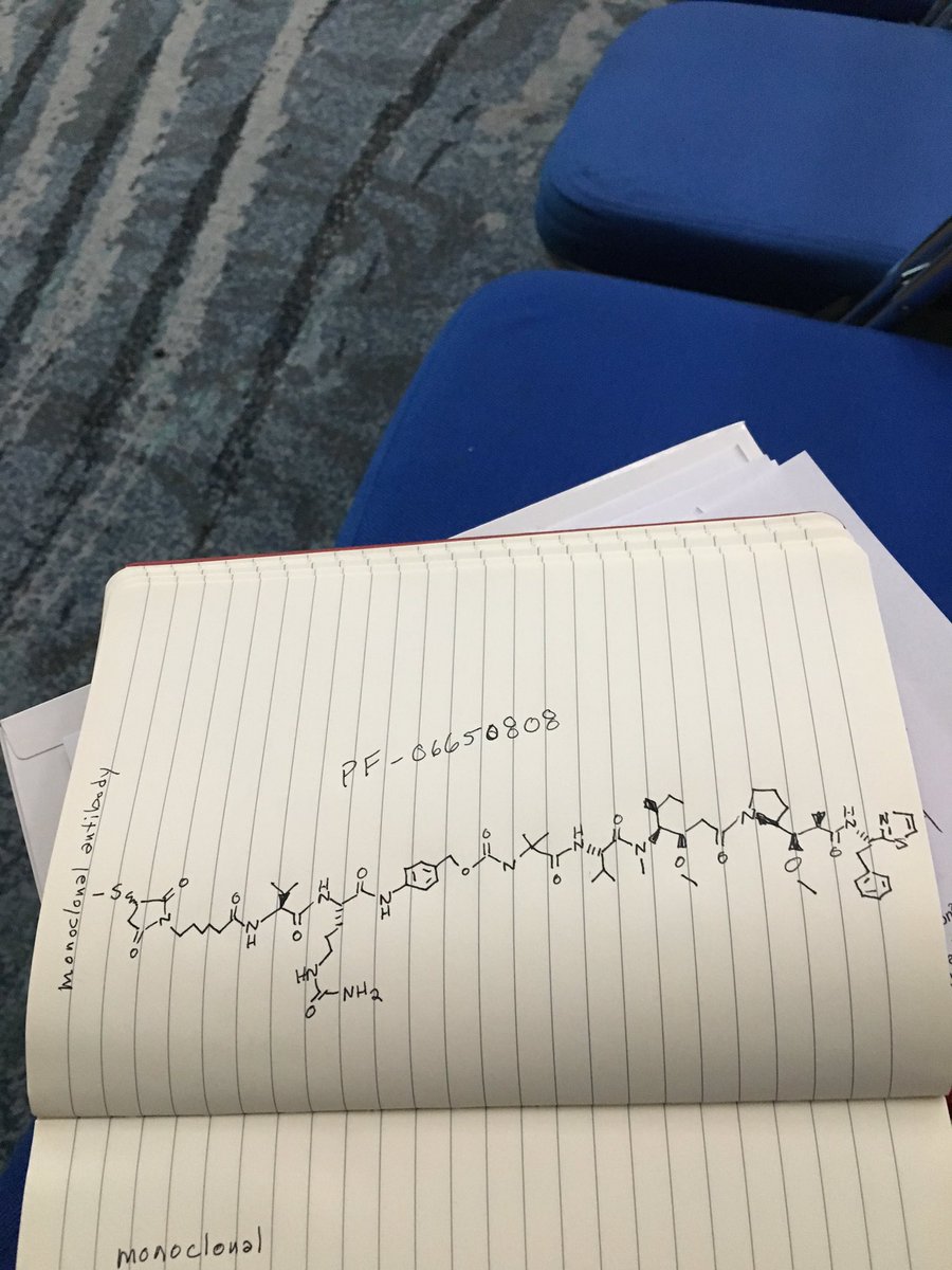
PF 06650808

Phase 1

compound inspired by auristatins

https://clinicaltrials.gov/ct2/show/NCT02129205

http://www.pfizer.com/sites/default/files/product-pipeline/8_7_2014_Pipeline_Update.pdf

ALL DATA COMING………

Notch-3 receptor antagonists

Neoplasms
Breast

Pfizer


Cancer

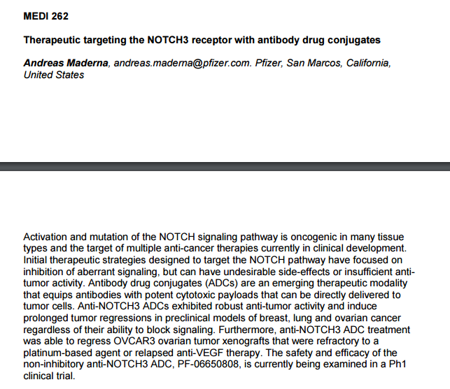
PF-06650808, is currently being examined in a Ph1 clinical trial (Protocol B7501001).

Notch3
Researchers are also exploring the use of Notch3 targeting. “The Notch pathway plays an important role in the growth of several solid tumours, including breast and ovarian cancer and melanoma,” explained Joerger. “In particular, Notch3 alterations such as gene amplification and upregulation are associated with poor patient survival. Research using Notch3 targeting as an innovative approach to treat solid malignancies included 27 patients unselected for Notch3 who received increasing doses of the anti-Notch3 antibody-drug conjugate PF-06650808. Responses were seen in two breast cancer patients (LBA 30). While preliminary, targeting Notch3 may become a new treatment approach in patients with selected solid tumours.”

The anti-Notch3 antibody-drug conjugate PF-06650808 is being developed by Pfizer.

  • 31 Jul 2014 Phase-I clinical trials in Solid tumours (Late-stage disease) in USA (Parenteral)
  • 30 Apr 2014 Preclinical trials in Solid tumours in USA (Parenteral)
  • 30 Apr 2014 Pfizer plans a phase I trial for Solid tumours (late-stage disease, second-line therapy or greater) in USA (NCT02129205)


251st Am Chem Soc (ACS) Natl Meet (March 13-17, San Diego) 2016, Abst MEDI 262


str1 STR2

/////////PF 06650808, PF-06650808, PF-6650808, monoclonal antibody, pfizer, phase 1, Solid tumours , Notch-3 receptor antagonists


C1(C(N(C(C1)=O)CCCCCC(=O)NC([C@H](C)C)C(=O)NC(C(=O)Nc2ccc(cc2)COC(=O)NC(C)(C)C(=O)N[C@@H](C(C)C)C(=O)[N@](C)C(C(CC)C)[C@@H](OC)CC(=O)N3CCC[C@H]3C(OO)C(C)C(=O)N[C@H](c4nccs4)CC)CCCNC(=O)N)=O)SC


DRUG APPROVALS BY DR ANTHONY MELVIN CRASTO …..FOR BLOG HOME CLICK HERE

Join me on Linkedin

View Anthony Melvin Crasto Ph.D's profile on LinkedIn

Join me on Facebook FACEBOOK

Join me on twitterFollow amcrasto on Twitter
Join me on google plus Googleplus

 amcrasto@gmail.com


P.S


THE VIEWS EXPRESSED ARE MY PERSONAL AND IN NO-WAY SUGGEST THE VIEWS OF THE PROFESSIONAL BODY OR THE COMPANY THAT I REPRESENT, amcrasto@gmail.com, +91 9323115463 India.

I , Dr A.M.Crasto is writing this blog to share the knowledge/views, after reading Scientific Journals/Articles/News Articles/Wikipedia. My views/comments are based on the results /conclusions by the authors(researchers). I do mention either the link or reference of the article(s) in my blog and hope those interested can read for details. I am briefly summarising the remarks or conclusions of the authors (researchers). If one believe that their intellectual property right /copyright is infringed by any content on this blog, please contact or leave message at below email address amcrasto@gmail.com. It will be removed ASAP

Share

Dipeptide-Based Metabolic Labeling of Bacterial Cells for Endogenous Antibody Recruitment

 MONOCLONAL ANTIBODIES  Comments Off on Dipeptide-Based Metabolic Labeling of Bacterial Cells for Endogenous Antibody Recruitment
Mar 232016
 
Abstract Image

The number of antibiotic-resistant bacterial infections has increased dramatically over the past decade. To combat these pathogens, novel antimicrobial strategies must be explored and developed. We previously reported a strategy based on hapten-modified cell wall analogues to induce recruitment of endogenous antibodies to bacterial cell surfaces. Cell surface remodeling using unnatural single d-amino acid cell wall analogues led to modification at the C-terminus of the peptidoglycan stem peptide. During peptidoglycan processing, installed hapten-displaying amino acids can be subsequently removed by cell wall enzymes. Herein, we disclose a two-step dipeptide peptidoglycan remodeling strategy aimed at introducing haptens at an alternative site within the stem peptide to improve retention and diminish removal by cell wall enzymes. Through this redesigned strategy, we determined size constraints of peptidoglycan remodeling and applied these constraints to attain hapten–linker conjugates that produced high levels of antibody recruitment to bacterial cell surfaces.

Dipeptide-Based Metabolic Labeling of Bacterial Cells for Endogenous Antibody Recruitment

Department of Chemistry, Lehigh University, Bethlehem, Pennsylvania 18015, United States
ACS Infect. Dis., Article ASAP
DOI: 10.1021/acsinfecdis.6b00007
Publication Date (Web): February 02, 2016
Copyright © 2016 American Chemical Society
*(M.M.P.) E-mail: map311@lehigh.edu.

ACS Editors’ Choice – This is an open access article published under an ACS AuthorChoice License, which permits copying and redistribution of the article or any adaptations for non-commercial purposes.

http://pubs.acs.org/doi/full/10.1021/acsinfecdis.6b00007

SEE OTHER PUBLICATIONS

Illumination of growth, division and secretion by metabolic …

A new metabolic cell-wall labelling method reveals peptidoglycan …

www.nature.com

Novel dipeptide PG labelling strategy.

Keywords:,  antibiotics,  bacterial,  surface,  remodeling,  d-amino acids, Dipeptide-Based Metabolic Labeling, Bacterial Cells,  Endogenous Antibody Recruitment

///

Share

FDA approves new treatment for inhalation anthrax, Anthim (obiltoxaximab) ETI-204

 MONOCLONAL ANTIBODIES  Comments Off on FDA approves new treatment for inhalation anthrax, Anthim (obiltoxaximab) ETI-204
Mar 222016
 

Anthim (obiltoxaximab)

ETI-204

March 21, 2016

On Friday, March 18, the U.S. Food and Drug Administration approved Anthim (obiltoxaximab) injection to treat inhalational anthrax in combination with appropriate antibacterial drugs. Anthim is also approved to prevent inhalational anthrax when alternative therapies are not available or not appropriate.

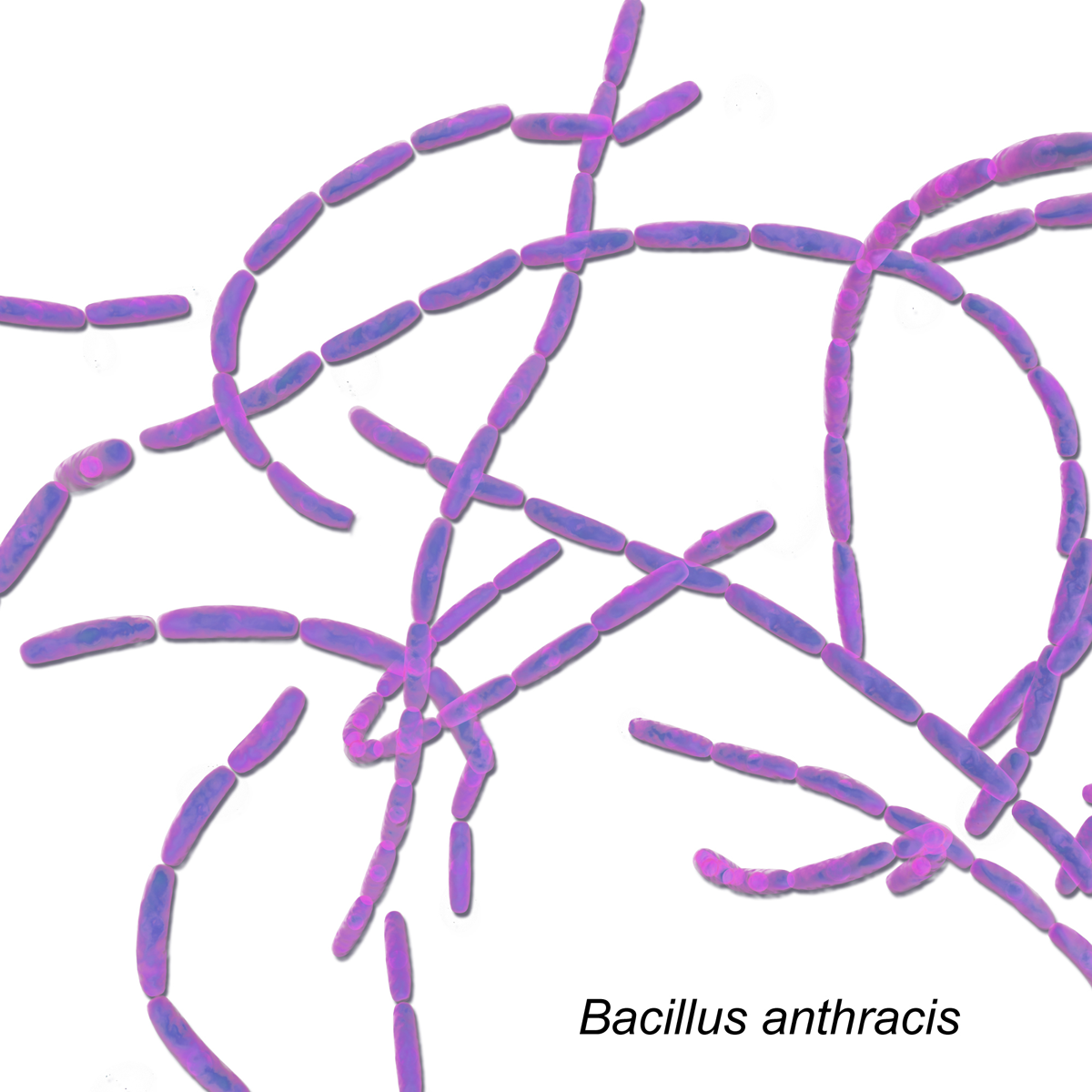
Inhalational anthrax is a rare disease that can occur after exposure to infected animals or contaminated animal products, or as a result of an intentional release of anthrax spores. It is caused by breathing in the spores of the bacterium Bacillus anthracis. When inhaled, the anthrax bacteria replicate in the body and produce toxins that can cause massive and irreversible tissue injury and death. Anthrax is a potential bioterrorism threat because the spores are resistant to destruction and can be spread by release in the air.

“As preparedness is a cornerstone of any bioterrorism response, we are pleased to see continued efforts to develop treatments for anthrax,” said Edward Cox, M.D., M.P.H, director of the Office of Antimicrobial Products in FDA’s Center for Drug Evaluation and Research.

Anthim is a monoclonal antibody that neutralizes toxins produced by B. anthracis. Anthim was approved under the FDA’s Animal Rule, which allows efficacy findings from adequate and well-controlled animal studies to support FDA approval when it is not feasible or ethical to conduct efficacy trials in humans.

Anthim’s effectiveness for treatment and prophylaxis of inhalational anthrax was demonstrated in studies conducted in animals based on survival at the end of the studies. More animals treated with Anthim lived compared to animals treated with placebo. Anthim administered in combination with antibacterial drugs resulted in higher survival outcomes than antibacterial therapy alone.

The safety of Anthim was evaluated in 320 healthy human volunteers. The most frequently reported side effects were headache, itching (pruritus), upper respiratory tract infections, cough, nasal congestion, hives, and bruising, swelling and pain at the infusion site.

Anthim carries a Boxed Warning alerting patients and health care providers that the drug can cause allergic reactions (hypersensitivity), including a severe reaction called anaphylaxis. Anthim should be administered in settings where patients can be monitored and treated for anaphylaxis. However, given that anthrax is a very serious and often deadly condition, the benefit of Anthim for treating anthrax is expected to outweigh this risk.

Anthim was developed by Elusys Therapeutics, Inc. of Pine Brook, New Jersey, in conjunction with the U.S. Department of Health and Human Services’ Biomedical Advanced Research and Development Authority.

Obiltoxaximab is a monoclonal antibody designed for the treatment of exposure to Bacillus anthracis spores (etiologic agent ofanthrax).[1]

This drug was developed by Elusys Therapeutics, Inc.

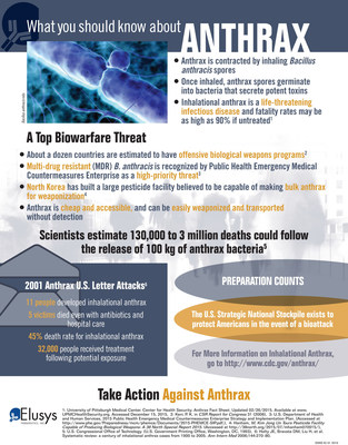
Infographic: What You Should Know About Anthrax


ANTHIM (obiltoxaximab) Data

The efficacy of ANTHIM for treatment and prophylaxis of inhalational anthrax was demonstrated in multiple studies in the cynomolgus macaque and NZW rabbit models of inhalational anthrax. These studies tested the efficacy of ANTHIM compared to placebo and the efficacy of ANTHIM in combination with antibacterial drugs relative to the antibacterial drugs alone. The primary endpoint was survival following challenge with B. anthracis.

Two studies in NZW rabbit and two studies in cynomolgus macaques evaluated treatment with ANTHIM 16mg/kg IV single dose compared to placebo in animals with systemic anthrax. Treatment with ANTHIM alone resulted in statistically significant improvement in survival relative to placebo in both species. Survival rates were 93% and 62% with ANTHIM compared to 0 placebo survivors in rabbits, and 47% and 31-35% survival with ANTHIM compared to 6% or 0% placebo survival in macaques.

ANTHIM administered in combination with antibacterial drugs (levofloxacin, ciprofloxacin and doxycycline) for the treatment of systemic inhalational anthrax disease resulted in higher survival outcomes than antibacterial therapy alone in multiple studies where ANTHIM and antibacterial therapy was given at various doses and treatment times.

ANTHIM administered as prophylaxis resulted in higher survival outcomes compared to placebo in multiple studies where treatment was given at various doses and treatment times. ANTHIM administered as prophylaxis resulted in higher survival outcomes compared to placebo in multiple studies where treatment was given at various doses and treatment times. In one study, cynomolgus macaques were administered ANTHIM 16 mg/kg at 18 hours, 24 hours or 36 hours after exposure. Survival was 6/6 (100%) at 18 hours, 5/6 (83%) at 24 hours, and 3/6 (50%) at 36 hours. Another cynomolgus macaque study evaluated ANTHIM 16 mg/kg administered 72, 48 or 24 hours prior to exposure. Survival was 100% at all three time points (14/14, 14/14, 15/15, respectively) at day 56 (end of study).

Elusys Therapeutics

Elusys Therapeutics, Inc., a private company based in Pine Brook, NJ, is focused on the development of antibody therapeutics for the treatment of infectious disease.

In November 2015, Elusys was awarded a $45M delivery order from the U.S. government to produce ANTHIM® for the U.S. Strategic National Stockpile (SNS), the U.S. government’s repository of critical medical supplies for public health emergency preparedness. Elusys has received grants and contracts from the USG totaling over $240 million to support ANTHIM’s development.

In March 2016, ANTHIM (obiltoxaximab) Injection, the company’s monoclonal antibody (mAb) anthrax antitoxin, received approval from the U.S. Food and Drug Administration (FDA) for the treatment of adult and pediatric patients with inhalational anthrax due toBacillus anthracis in combination with appropriate antibacterial drugs, and for prophylaxis of inhalational anthrax due to B. anthracis when alternative therapies are not available or not appropriate. ANTHIM should only be used for prophylaxis when its benefit for prevention of inhalational anthrax outweighs the risk of hypersensitivity and anaphylaxis. The effectiveness of ANTHIM is based solely on efficacy studies in animal models of inhalational anthrax. There have been no studies of the safety or pharmacokinetics (PK) of ANTHIM in the pediatric population. Dosing in pediatric patients was derived using a population PK approach. ANTHIM does not have direct antibacterial activity. ANTHIM should be used in combination with appropriate antibacterial drugs. ANTHIM is not expected to cross the blood-brain barrier and does not prevent or treat meningitis.


Company Elusys Therapeutics Inc.
Description High-affinity humanized mAb against the Bacillus anthracis protective antigen that inhibits binding of anthrax toxins
Molecular Target Bacillus anthracis protective antigen
Mechanism of Action Antibody
Therapeutic Modality Biologic: Antibody
Latest Stage of Development Approved
Standard Indication Anthrax
Indication Details Treat and prevent anthrax infection; Treat anthrax infection
Regulatory Designation U.S. – Fast Track (Treat and prevent anthrax infection);
U.S. – Orphan Drug (Treat and prevent anthrax infection)

///////////Anthim, obiltoxaximab, fda 2016, Orphan Drug,


Share

A human recombinant monoclonal antibody to cocaine: Preparation, characterization and behavioral studies

 MONOCLONAL ANTIBODIES  Comments Off on A human recombinant monoclonal antibody to cocaine: Preparation, characterization and behavioral studies
Sep 232014
 

image


A human recombinant monoclonal antibody to cocaine: Preparation, characterization and behavioral studies

CLICK…….http://www.sciencedirect.com/science/article/pii/S0960894X1400866X

Volume 24, Issue 19, 1 October 2014, Pages 4664–4666

DOI: 10.1016/j.bmcl.2014.08.035
Lisa M. Eubanks, Beverly A. Ellis, Xiaoqing Cai, Joel E. Schlosburg, Kim D. Janda

Abstract

Cocaine abuse remains prevalent worldwide and continues to be a major health concern; nonetheless, there is no effective therapy. Immunopharmacotherapy has emerged as a promising treatment strategy by which anti-cocaine antibodies bind to the drug blunting its effects. Previous passive immunization studies using our human monoclonal antibody, GNCgzk, resulted in protection against cocaine overdose and acute toxicity. To further realize the clinical potential of this antibody, a recombinant IgG form of the antibody has been produced in mammalian cells. This antibody displayed a high binding affinity for cocaine (low nanomolar) in line with the superior attributes of the GNCgzk antibody and reduced cocaine-induced ataxia in a cocaine overdose model.


Keywords

  • Cocaine;
  • Overdose;
  • Immunopharmacotherapy;
  • Passive immunization;
  • Monoclonal antibody
Share

FDA approves Keytruda for advanced melanoma, First PD-1 blocking drug to receive agency approval

 MONOCLONAL ANTIBODIES, Uncategorized  Comments Off on FDA approves Keytruda for advanced melanoma, First PD-1 blocking drug to receive agency approval
Sep 052014
 

September 4, 2014

FDA Release

The U.S. Food and Drug Administration today granted accelerated approval to Keytruda (pembrolizumab) for treatment of patients with advanced or unresectable melanoma who are no longer responding to other drugs.

Melanoma, which accounts for approximately 5 percent of all new cancers in the United States, occurs when cancer cells form in skin cells that make the pigment responsible for color in the skin. According to the National Cancer Institute, an estimated 76,100 Americans will be diagnosed with melanoma and 9,710 will die from the disease this year.

Keytruda is the first approved drug that blocks a cellular pathway known as PD-1, which restricts the body’s immune system from attacking melanoma cells. Keytruda is intended for use following treatment with ipilimumab, a type of immunotherapy. For melanoma patients whose tumors express a gene mutation called BRAF V600, Keytruda is intended for use after treatment with ipilimumab and a BRAF inhibitor, a therapy that blocks activity of BRAF gene mutations.

“Keytruda is the sixth new melanoma treatment approved since 2011, a result of promising advances in melanoma research,” said Richard Pazdur, M.D., director of the Office of Hematology and Oncology Products in the FDA’s Center for Drug Evaluation and Research. “Many of these treatments have different mechanisms of action and bring new options to patients with melanoma.”

The five prior FDA approvals for melanoma include: ipilimumab (2011), peginterferon alfa-2b (2011), vemurafenib (2011), dabrafenib (2013), and trametinib (2013).

The FDA granted Keytruda breakthrough therapy designation because the sponsor demonstrated through preliminary clinical evidence that the drug may offer a substantial improvement over available therapies. It also received priority review and orphan product designation. Priority review is granted to drugs that have the potential, at the time the application was submitted, to be a significant improvement in safety or effectiveness in the treatment of a serious condition. Orphan product designation is given to drugs intended to treat rare diseases.

The FDA action was taken under the agency’s accelerated approval program, which allows approval of a drug to treat a serious or life-threatening disease based on clinical data showing the drug has an effect on a surrogate endpoint reasonably likely to predict clinical benefit to patients. This program provides earlier patient access to promising new drugs while the company conducts confirmatory clinical trials. An improvement in survival or disease-related symptoms has not yet been established.

Keytruda’s efficacy was established in 173 clinical trial participants with advanced melanoma whose disease progressed after prior treatment. All participants were treated with Keytruda, either at the recommended dose of 2 milligrams per kilogram (mg/kg) or at a higher dose of 10 mg/kg. In the half of the participants who received Keytruda at the recommended dose of 2 mg/kg, approximately 24 percent had their tumors shrink. This effect lasted at least 1.4 to 8.5 months and continued beyond this period in most patients. A similar percentage of patients had their tumor shrink at the 10 mg/kg dose.

Keytruda’s safety was established in the trial population of 411 participants with advanced melanoma. The most common side effects of Keytruda were fatigue, cough, nausea, itchy skin (pruritus), rash, decreased appetite, constipation, joint pain (arthralgia) and diarrhea. Keytruda also has the potential for severe immune-mediated side effects. In the 411 participants with advanced melanoma, severe immune-mediated side effects involving healthy organs, including the lung, colon, hormone-producing glands and liver, occurred uncommonly.

Keytruda is marketed by Merck & Co., based in Whitehouse Station, New Jersey.


Pembrolizumab, LambrolizumabMK-3475

STRUCTURAL FORMULA
Heavy chain
QVQLVQSGVE VKKPGASVKV SCKASGYTFT NYYMYWVRQA PGQGLEWMGG 50
INPSNGGTNF NEKFKNRVTL TTDSSTTTAY MELKSLQFDD TAVYYCARRD 100
YRFDMGFDYW GQGTTVTVSS ASTKGPSVFP LAPCSRSTSE STAALGCLVK 150
DYFPEPVTVS WNSGALTSGV HTFPAVLQSS GLYSLSSVVT VPSSSLGTKT 200
YTCNVDHKPS NTKVDKRVES KYGPPCPPCP APEFLGGPSV FLFPPKPKDT 250
LMISRTPEVT CVVVDVSQED PEVQFNWYVD GVEVHNAKTK PREEQFNSTY 300
RVVSVLTVLH QDWLNGKEYK CKVSNKGLPS SIEKTISKAK GQPREPQVYT 350
LPPSQEEMTK NQVSLTCLVK GFYPSDIAVE WESNGQPENN YKTTPPVLDS 400
DGSFFLYSRL TVDKSRWQEG NVFSCSVMHE ALHNHYTQKS LSLSLGK 447
Light chain
EIVLTQSPAT LSLSPGERAT LSCRASKGVS TSGYSYLHWY QQKPGQAPRL 50′
LIYLASYLES GVPARFSGSG SGTDFTLTIS SLEPEDFAVY YCQHSRDLPL 100′
TFGGGTKVEI KRTVAAPSVF IFPPSDEQLK SGTASVVCLL NNFYPREAKV 150′
QWKVDNALQS GNSQESVTEQ DSKDSTYSLS STLTLSKADY EKHKVYACEV 200′
THQGLSSPVT KSFNRGEC 218′
Disulfide bridges
22-96 22”-96” 23′-92′ 23”’-92”’ 134-218′ 134”-218”’ 138′-198′ 138”’-198”’
147-203 147”-203” 226-226” 229-229” 261-321 261”-321” 367-425 367”-425”
Glycosylation sites (N)
Asn-297 Asn-297”
lambrolizumab, or MK-3475

1374853-91-4

C6504H10004N1716O2036S46 (peptide)
MOL. MASS 146.3 kDa (peptide)

Pembrolizumab, Lambrolizumab (also known as MK-3475) is a drug in development by Merck that targets the PD-1 receptor. The drug is intended for use in treating metastatic melanoma.

http://www.ama-assn.org/resources/doc/usan/lambrolizumab.pdf  structureof lambrolizumab, or MK-3475

https://download.ama-assn.org/resources/doc/usan/x-pub/pembrolizumab.pdf  

Statement on a Nonproprietary Name Adopted by the USAN Council. November 27, 2013.

see above link for change in name

may 2, 2013,

An experimental drug from Merck that unleashes the body’s immune system significantly shrank tumors in 38 percent of patients with advanced melanoma, putting the company squarely in the race to bring to market one of what many experts view as the most promising class of drugs in years.

The drugs are attracting attention here at the annual meeting of the American Society of Clinical Oncology, even though they are still in the early stage of testing. Data from drugs developed by Bristol-Myers Squibb and by Roche had already been released.

The drugs work by disabling a brake that prevents the immune system from attacking cancer cells. The brake is a protein on immune system cells called programmed death 1 receptor, or PD-1.

Merck’s study, which was presented here Sunday and also published in the New England Journal of Medicine, involved 135 patients. While tumors shrank in 38 percent of the patients over all, the rate was 52 percent for patients who got the highest dose of the drug, which is called lambrolizumab, or MK-3475.

But that is what is disclosed tonight, as to pembrolizumab, or MK-3475. Wow. With over $44 billion in 2013 worldwide revenue, that disclosure implies (to seasoned SEC lawyers) that spending on this one drug (or, biologic, to be more technical about it — but remember 40 years ago, Merck had no protein chain biologics research & development programs in its pipe — only chemical drug compounds). . . is material, to that number. Normally that would, in turn, mean that the spending is approaching 5 per cent of revenue. So — Merck may be spending $2.2 billion over the next 12 rolling months, on MK-3475. That’s one BIGhairy science bet, given that Whitehouse Station likely already had over $2 billion invested in the program, at year end 2013.

About Pembrolizumab
Pembrolizumab (MK-3475) is an investigational selective, humanized monoclonal anti-PD-1 antibody designed to block the interaction of PD-1 on T-cells with its ligands, PD-L1 and PD-L2, to reactivate anti-tumor immunity. Pembrolizumab exerts dual ligand blockade of PD-1 pathway.
Today, pembrolizumab is being evaluated across more than 30 types of cancers, as monotherapy and in combination. It is anticipated that by the end of 2014, the pembrolizumab development program will grow to more than 24 clinical trials across 30 different tumor types, enrolling an estimated 6,000 patients at nearly 300 clinical trial sites worldwide, including new Phase 3 studies in head and neck and other cancers. For information about Merck’s oncology clinical studies, please click here.
The Biologics License Application (BLA) for pembrolizumab is under priority review with the U.S. Food and Drug Administration (FDA) for the proposed indication for the treatment of patients with advanced melanoma previously-treated with ipilimumab; the PDUFA date is October 28, 2014. Pembrolizumab has been granted FDA’s Breakthrough Therapy designation for advanced melanoma. If approved by the FDA, pembrolizumab has the potential to be the first PD-1 immune checkpoint modulator approved in this class. The company plans to file a Marketing Authorization Application in Europe for pembrolizumab for advanced melanoma in 2014.
About Head and Neck Cancer
Head and neck cancers are a related group of cancers that involve the oral cavity, pharynx and larynx. Most head and neck cancers are squamous cell carcinomas that begin in the flat, squamous cells that make up the thin surface layer (epithelium) of the head and neck (called the). The leading risk factors for head and neck cancer include tobacco and alcohol use. Infection with certain types of HPV, also called human papillomaviruses, is a risk factor for some types of head and neck cancer, specifically cancer of the oropharynx, which is the middle part of the throat including the soft palate, the base of the tongue, and the tonsils. Each year there are approximately 400,000 cases of cancer of the oral cavity and pharynx, with 160,000 cancers of the larynx, resulting in approximately 300,000 deaths.


About Merck Oncology: A Focus on Immuno-Oncology
At Merck Oncology, our goal is to translate breakthrough science into biomedical innovations to help people with cancer worldwide. Harnessing immune mechanisms to fight cancer is the priority focus of our oncology research and development program. The Company is advancing a pipeline of immunotherapy candidates and combination regimens. Cancer is one of the world’s most urgent unmet medical needs. Helping to empower people to fight cancer is our passion. For information about Merck’s commitment to Oncology visit the Oncology Information Center at http://www.mercknewsroom.com/oncology-infocenter.


About Merck
Today’s Merck is a global healthcare leader working to help the world be well. Merck is known as MSD outside the United States and Canada. Through our prescription medicines, vaccines, biologic therapies, and consumer care and animal health products, we work with customers and operate in more than 140 countries to deliver innovative health solutions. We also demonstrate our commitment to increasing access to healthcare through far-reaching policies, programs and partnerships. For more information, visit www.merck.com and connect with us on Twitter, Facebook and YouTube.


Hamid, O; Robert, C; Daud, A; Hodi, F. S.; Hwu, W. J.; Kefford, R; Wolchok, J. D.; Hersey, P; Joseph, R. W.; Weber, J. S.; Dronca, R; Gangadhar, T. C.; Patnaik, A; Zarour, H; Joshua, A. M.; Gergich, K; Elassaiss-Schaap, J; Algazi, A; Mateus, C; Boasberg, P; Tumeh, P. C.; Chmielowski, B; Ebbinghaus, S. W.; Li, X. N.; Kang, S. P.; Ribas, A (2013). “Safety and tumor responses with lambrolizumab (anti-PD-1) in melanoma”. New England Journal of Medicine 369 (2): 134–44. doi:10.1056/NEJMoa1305133PMID 23724846

key words
FDA,  approved,  Keytruda,  advanced melanoma, PD-1 blocking drug, pembrolizumab, LambrolizumabMK-3475, Monoclonal antibody


Share

Large Scale Production Of MAbs

 MONOCLONAL ANTIBODIES  Comments Off on Large Scale Production Of MAbs
Jun 132014
 

Production of MAb

Fig.1 Production of MAb

Large Scale Production Of MAbs:

Commercially, on large scale, MAbs are produced by two methods.

(a) Ascites production in mice

(b) In-vitro fermentation

The production method is summarized in Fig.no.2a & 2b.

a) Ascites Production In Mice:

The first monoclonal antibodies approved by FDA for therapeutic use OKTS, is produced by ascitic technology19.

In this method hybridoma cells are injected into peritoneal cavity of histocompatible mice. The mice are pretreated by i.p. injection of Pristane to irritate the peritoneal cavity which facilitates the growth of ascitic tumor. The fluid produced may contain the high concentration of secreted MAbs, 2 to 20 μg / ml and 2 to 6 ml or more can be harvested per mouse. Comparison of different MAb production22,23 methods is shown inTable 1.

Drawbacks of this method are:

1. It is very costly, very difficult and not reliable.

2. Product may get contaminated with mouse immunoglobulins and also with other mouse proteins.

3. Viruses can be introduced as contaminants.

4. Antibody yield is often less as compared to other methods.

b) In-Vitro Fermentation:

In this method, the cells are grown and gradually moved to larger and larger culture ensuring exponential growth. Typical antibody levels in the culture supernatant ranges from 5-50 μg/ml depending on the individual clone and on cell density. When more production of antibody is required 1-litre cultures in roller bottles are used. Required cells are removed from rest of media by centrifugation or filtration, generally followed by ultra filtration step for concentrating the filtrate by up to 20 folds.

Advantages of this method are:

(1) As serum required in culture media is reduced, it is cost effective.

(2) There will not be any contamination with mouse immunoglobulin.

But the major drawback is that of contamination of final product with serum or protein based growth factors.

Table 1: Comparison of different MAb production methods.

Production system

Scale

Volume (ml)

Concentration (mg/ml)

Production time (weeks)

Quality

Ascites (in vivo)

20-250 mg

5-10

< 20

2-3

Low
Stir growth

100-2500

0.01-0.1

2-3

High
Dialysis membrane

< 50 mg

10-25

0.1-1.5

2-5

High
Roller bottles

< 2 gm

100-2000

0.01-0.2

2-6

High
Hollow fiber

0.15-30 gm

25-1000

0.2-0.3

3-12

High
Fermentor

2-100 gm

< 2000 lit

0.05-0.5

2-12

High

 MAb Production

Fig. 2a:  MAb Production (Flowchart)

 Freeze Dried MAb Production

Fig. 2b:  Freeze Dried MAb Production (Flowchart)

i) Purification:

Contamination, during production process, such as protein, nucleic acid, endotoxins, immunoglobulin and adventitious agent can be removed by purification method. The purification methods such as precipitation with ammonium sulphate, zone electrophoresis, ion exchange chromatography, hydrophobic interaction chromatography, gel filtration and affinity chromatography are used19.

· Affinity chromatography is often used for initial purification.

· Ion exchange chromatography is used for removing endotoxins and DNA.

· Gel filtration chromatography can remove both high and low molecular form of monoclonal antibodies and it is usually used as the final polishing step.

j) Characterization:

The final determination of monoclonality requires biochemical and biophysical characterization of the immunoglobulin. It is also characterized immunochemically to define its affinity for antigen, its immunoglobulin subclass, the epitopes for which it is specific and the effective number of binding site that it possesses19.

k) Final Processing:

Depending upon the intended application, the antibody may be conjugated to specific radionuclide or toxin. Then the stabilizing agent is added, and the product is filled into final container under inert gas or other specialized conditions.  Lyophillization is frequently applied to get freeze dried product.

Antigenicity Of Murine MAb:

The main problem for mouse MAb is that, human body recognizes it as a foreign agent and produces antibodies against such mouse MAb. The induced human anti-mouse antibodies (HAMA) quickly reduce the effectiveness of mouse MAb and also their interaction may lead to allergic reactions.

To overcome the problem, Human MAbs can be used. Though difficult, this is possible by fusion of EBV (Epstein Barr Virus) transformed human B-lymphocyte with appropriate fusion partners21. EBV is a lymphotrophic DNA herpes virus which is capable of converting normal B-lymphocytes of human and/or mouse into cancer cell having proliferating capacity in vitro. But the presence of EBV as contaminant can pose a problem of producing cancer24.

Even the human-human hybridomas producing MAbs have been produced 25,26. Olsson and Kaplan in the year 1980 produced first human-human myeloma (SKO-007), against the hapten 2, 4-dinitrophenyl (DNP) 19.

The routine production of human MAbs is prevented due to following reason:-

  • Sources of antibody producing cells27.
  • Reliable methods for lymphocytes immortalization.
  • Stability28 and antibody producing capacity.
  • Administration of some antigens to humans could endanger their health29.
  • Recovery of B-lymphocytes from the spleen of human is impracticable.
  • The fusion of human lymphocytes with human lymphoblastoid cell lines is a very inefficient process.
  • Low production yield of human monoclonal antibody.

Hence, other alternatives methods come forth.

Advantages Of MAbs:

  • Pure one molecular species with high specificity for a particular antigenic target.
  • Anti-serum titer values are high.
  • Antibodies with high avaidity can be produced.
  • In vitro and in vivo production is possible.
  • Radiolabelling and fluorescent conjugation of monoclonal antibody are easy.

Disadvantages Of MAbs:

  • Initial cost involved in the technique is high. However, continuous production is somewhat economical.
  • Methods are time consuming.
  • Antigenicity of Murine MAb.
  • MAbs have comparatively less complement fixing ability than that of convectional antiserum.
  • MAbs are highly selective for a particular single antigenic determinant. This renders them incapable of distinguish between different molecules, cells bearing the chemical structure or determinants except one against which it is targeted.
  • The high antibody avidity (energy of binding to an antigen) of MAbs is advantageous for immunoassay but some property is undesirable for purification process.
Share
Follow

Get every new post on this blog delivered to your Inbox.

Join other followers: