AUTHOR OF THIS BLOG

DR ANTHONY MELVIN CRASTO, WORLDDRUGTRACKER

DR ANTHONY MELVIN CRASTO Ph.D

DR ANTHONY MELVIN CRASTO, Worlddrugtracker, Born in Mumbai in 1964 and graduated from Mumbai University, Completed his PhD from ICT ,1991, Mumbai, India, in Organic chemistry, The thesis topic was Synthesis of Novel Pyrethroid Analogues, Currently he is working with AFRICURE PHARMA as ADVISOR earlier GLENMARK LS Research centre as consultant,Principal Scientist, Process Research (bulk actives) at Mahape, Navi Mumbai, India. Prior to joining Glenmark, he worked with major multinationals like Hoechst Marion Roussel, now sSanofi, Searle India ltd, now Rpg lifesciences, etc. he is now helping millions, has million hits on google on all organic chemistry websites. His New Drug Approvals, Green Chemistry International, Eurekamoments in organic chemistry are some most read blogs He has hands on experience in initiation and developing novel routes for drug molecules and implementation them on commercial scale over a 32 year tenure, good knowledge of IPM, GMP, Regulatory aspects, he has several international drug patents published worldwide . He gas good proficiency in Technology transfer, Spectroscopy, Stereochemistry, Synthesis, polymorphism etc He suffered a paralytic stroke in dec 2007 and is bound to a wheelchair, this seems to have injected feul in him to help chemists around the world, he is more active than before and is pushing boundaries, he has one lakh connections on all networking sites, He makes himself available to all, contact him on +91 9323115463, amcrasto@gmail.com

PDE4 inhibitor , Sumitomo Dainippon Pharma Company

 Uncategorized  Comments Off on PDE4 inhibitor , Sumitomo Dainippon Pharma Company
May 192016
 

Figure


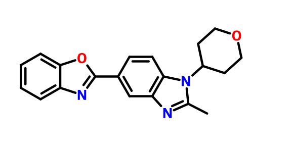
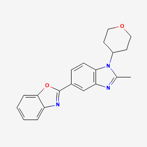
2-[2-Methyl-1-(tetrahydro-2H-pyran-4-yl)-1H-benzimidazol-5-yl]-1,3-benzoxazole Hemifumarate

Sumitomo Dainippon Pharma Company,

STR1

SCHEMBL2688684.png

CAS FREE FORM 1256966-65-0

Benzoxazole, 2-​[2-​methyl-​1-​(tetrahydro-​2H-​pyran-​4-​yl)​-​1H-​benzimidazol-​5-​yl]​-

MF C20 H19 N3 O2, MW, 333.38 FREE FORM
NMR FOR HEMIFUMARATE

1H NMR (400 MHz, DMSO-d6)

δ 13.1 (br, 1H), 8.33 (d, J = 1.5 HZ, 1H), 8.06 (dd, J = 5.1, 1.6 Hz, 1H), 7.89 (d, J = 0.8 Hz, 1H), 7.82–7.76 (m, 2H), 7.43–7.38 (m, 2H), 6.64 (s, 1H), 4.71–4.62 (m, 1H), 4.06 (dd, J = 11.4, 4.3 Hz, 2H), 3.58 (dd, J = 11.7, 11.4 Hz, 2H), 2.67 (s, 3H), 2.47–2.36 (m, 2H), 1.90–1.86 (m, 2H).

13C NMR (100 MHz, DMSO-d6)

δ 165.92, 163.26, 153.94, 150.20, 142.94, 141.75, 136.21, 133.93, 124.94, 124.67, 120.89, 119.40, 117.70, 112.44, 110.72, 66.50, 52.67, 30.70, 14.62.
Compound 1 is a PDE4 inhibitor and is expected to improve memory impairment. In addition to the mechanism of action, 1 enhances BDNF signal transduction and induces NXF, a brain specific transcription factor, in the presence of low concentrations of BDNF. NXF induction is expected to lead to nerve regeneration and neuroprotective efficacy.
US88290352014-09-09Agent for treatment or prevention of diseases associated with activity of neurotrophic factors
 STR1
Example 11
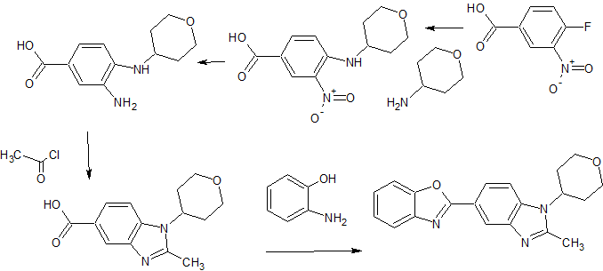
5- (benzoxazol-2-yl) -2-methyl -1-(tetrahydropyran-4-yl) benzimidazole  eggplant flask (100 mL), 2- methyl-1- (tetrahydropyran – 4-yl) reference benzimidazole-5-carboxylic acid (example 4-3) (0.64 g, 2.46 mmol ), 2- amino-phenol (0.32 g, 2.95 mmol), and polyphosphoric acid (about 18 g) put, heated to 160 ℃, and the mixture was stirred for 17 hours. After cooling, ice was added, and the mixture was about pH 9 the liquid with concentrated aqueous ammonia (28%). Extraction with chloroform (about 50 mL X 3 times), dried over anhydrous magnesium sulfate, the crude product obtained by distilling off the solvent (0.08 g) PTLC (CHCl 3 by weight deploy purified), the title compound ( 0.002 g, 0.2% yield) was obtained as a yellow-brown semi-solid. 1H-NMR (CDCl 3 ) Deruta (Ppm): 1.88-1.92 (M, 2 H), 2.58-2.68 (M, 2 H), 2.70 (S, 3 H), 3.57-3.64 (M , 2 H), 4.21-4.25 (m , 2 H), 4.43-4.49 (m, 1 H), 7.29 (d, 1H, J = 9.2 Hz), 7.33-7.35 (m, 2 H ), 7.59-7.62 (m, 1 H ), 7.76-7.78 (m, 1 H), 8.18 (dd, 1 H, J = 8.6, 1.6 Hz), 8.57 (d, 1 H, J = 1.4 Hz).

PAPER

Abstract Image

A short and practical synthetic route of a PDE4 inhibitor (1) was established by using Pd–Cu-catalyzed C–H/C–Br coupling of benzoxazole with a heteroaryl bromide. The combination of Pd(OAc)2-Cu(OTf)2-PPh3 was found to be effective for this key step. Furthermore, telescoping methods were adopted to improve the yield and manufacturing time, and a two-step synthesis of1 was accomplished in 71% overall yield.

Direct Synthesis of a PDE4 Inhibitor by Using Pd–Cu-Catalyzed C–H/C–Br Coupling of Benzoxazole with a Heteroaryl Bromide

Process Chemistry Research and Development Laboratories, Technology Research & Development Division andDSP Cancer Institute, Sumitomo Dainippon Pharma Company, Ltd., 3-1-98 Kasugade-naka, Konohana-ku, Osaka 554-0022, Japan
Org. Process Res. Dev., Article ASAP
DOI: 10.1021/acs.oprd.6b00106

///////////PDE4 inhibitor , Sumitomo Dainippon Pharma Company

Cc1nc3cc(ccc3n1C2CCOCC2)c4nc5ccccc5o4

Share

ICH M7

 regulatory, Uncategorized  Comments Off on ICH M7
May 192016
 

ICH M7


Although relatively quiet in terms of any specific regulatory activities, the last 6 months have seen a plethora of publications that are associated with the ICH M7 guideline. Prominent within these was the Special Edition of Organic Process Research & Development in November 2015. This special edition focused on mutagenic impurities, examining the challenges and also opportunities faced when seeking to implement ICH M7.(5) This was timely as it aligned with the effective date for ICH M7 of January 2016; the guideline when finalized in June 2014 having a defined implementation phase of 18 months. ICH M7 is, in general, a well-written guideline that provides a flexible and pragmatic framework by which the risk posed by mutagenic impurities can be effectively managed. The flexibility provided by the guideline and the opportunities this presents in terms of science and risk based thinking are examined in depth through a series of articles within the special edition.
A tabulated summary of the special edition is described in Table 1.

Table 1

subject highlights authors
Is Avoidance of Genotoxic Intermediates/Impurities Tenable for Complex, Multistep Syntheses? A survey of over 300 synthetic publications in OPR&D over a 10 year period clearly demonstrated that the synthesis of synthetic APIs was untenable without the use reactive, potentially mutagenic reagents/intermediates. That the principle of avoidance was fundamentally flawed Elder, D. P.; Teasdale, A.(6)
Strategies To Address Mutagenic Impurities Derived from Degradation in Drug Substances and Drug Products The paper outlines a strategy for the systematic assessment of the risk posed by mutagenic degradants, describing how this relates to stress testing and long-term stability studies. Within this it seeks to define appropriate thresholds for identification directly related to the extent of degradation Kleinman, M. H.; Teasdale, A.; Baertschi, S. W. et al.(7)
Assessing the Risk of Potential Genotoxic Degradation Products in a Small Molecule Kinase Inhibitor Drug Substance and Drug Product The degradation profile resulting from stress testing of galunisertib is described, focusing on formation of two N-oxides, examining the site of oxidation and the relevance of the pathway under typical storage conditions. Strege, M. A.; Osborne, L. M.; Hetrick, E. M. et al.(8)
Mutagenic Alkyl-Sulfonate Impurities in Sulfonic Acid Salts: Reviewing the Evidence and Challenging Regulatory Perceptions Provides a comprehensive review of the existing evidence relating to sulfonate esters, examining the comprehensive mechanistic and kinetic studies and safety data. It also examines the current regulatory approaches and how this appears misaligned with the data. Snodin, D.; Teasdale, A.(9)
Mutagenic Impurities: Precompetitive Collaborative and Data Sharing Initiatives Examines the nature, impact, and successes of a series of cross industry initiatives covering areas such as structural evaluation (Q)SAR, data sharing–aromatic amines, boronic acids, purging and degradation. Elder, D. P.; Williams, R.; Harvey et al.(10)
Do Carboxylic/Sulfonic Acid Halides Really Present a Mutagenic and Carcinogenic Risk As Impurities in Final Drug Products? Examines evidence that indicates that in the case of both sulfonyl and acyl chlorides that Ames positive results relate to generation of a reactive species, halodimethyl sulphides (HDMSs) through reaction with DMSO and that this is the root cause of a positive response. Confirmatory negative data from other test solvents is also provided Amberg, A.; Harvey, J.; Spirkl, H.-P. et al.(11)
Boronic Acids and Derivatives—Probing the Structure–Activity Relationships for Mutagenicity The primary purpose is to raise awareness of the potentially mutagenic nature of boronic acids and stimulate further discussion/research in the areas. It provides mutagenicity data for some 40+ examples, examining the current status of in silico predictions and postulates a potential mechanism related to oxidation of boronic acids to yield oxygen radicals Hansen, M. H.; Jolly, R. A.; Linder, R. J.(12)
A Kinetics-Based Approach for the Assignment of Reactivity Purge Factors Details an experimental approach that utilizes kinetic analysis to facilitate assignment of reactivity purge values. Betori, R. C.; Kallemeyn, J. M.; Welch, D. S.(13)
A Generic Industry Approach to Demonstrate Efficient Purification of Potential Mutagenic Impurities (PMIs) in the Synthesis of Drug Substances Based on vortioxetine and its associated PMIs predicted purge values based on the system described by Teasdale et al.(15) are compared with experimental values. The results show good correlation concluding that theoretical purge values can be used to predict purging of PMIs. Lapanja N, Zupanĉiĉ B, Toplak Ĉasar R et al(14)
Evaluation and Control of Mutagenic Impurities in a Development Compound: Purge Factor Estimates versus Measured Amounts The purging of MIs associated with the synthesis of MK-8876 were assessed using the approach described by Teasdale et al.(15)These predicted values were compared to measured values and shown to be conservative in comparison to experimental data. McLaughlin, M.; Dermenijan, R. K.; Jin, Y. et al.(16)
Several papers focused on control options, specifically ICH option 4, involving evaluation of the impact of process conditions upon the purging of mutagenic impurities. This concept was first described by Teasdale et al. in 2010(17) and augmented by a cross-industry evaluation published in 2013.(15) The practical use of such tools is examined through two papers, that of Nevenka et al.(14) and McLaughlin et al.(16) This is augmented by a further publication by Welch et al.(13)that describes work now being undertaken by an industry consortium to develop this tool still further as a robust in silico tool (Mirabilis). Welch et al. describe the work being undertaken to fully evaluate the potential fate of MIs under a range of common chemical transformations. A critical finding of these studies, examined through the reaction of benzyl bromide with triethylamine, was alignment between the rate constants and half-lives of the reaction of benzyl bromide with triethylamine in isolation and as a low-level impurity in the TBS protection of benzyl alcohol (Figure 2). This established the proof of concept that the kinetic information obtained from the stand-alone reaction can be used to predict impurity conversion in a more complex reaction.

Figure

Figure 2. Alignment between the reaction of benzyl bromide with triethylamine in isolation and as a low-level impurity in the TBS protection of benzyl alcohol.

Another area addressed in the special edition is that of sulfonate esters. This relates to the use of a sulfonic acid, used to form an API salt and the potential formation of sulfonate esters through reaction with alcoholic solvents. Snodin and Teasdale(9) have reviewed the available literature information concluding that the extensive evidence supports the view that such concerns are grossly exaggerated. In parallel to this publication there have been a series of correspondences involving the EMA quality working party, the following points were released following discussion at the CVMP committee.(18)

“The Committee endorsed the QWP response to the EDQM request for an opinion on new information on alkyl sulfonates. The QWP reviewed the article from Snodin et al. QWP acknowledges the scientific rationale in this article and that the formation of alkyl sulfonates is very low and very much depends on the reaction conditions. This makes the presence of these mutagenic impurities at toxicologically significant levels unlikely. However, as the presence and formation of these alkyl sulfonates cannot be totally excluded, QWP proposes the following approach: marketing authorization holders should justify via Risk Assessment that alkyl sulfonates are not expected to be present for their product, which may be sufficient.”

Of concern within this text is the comment that the presence and formation cannot be totally excluded; this is despite the evidence pointing clearly to fact that it can.

Similarly at the end of February EDQM issued a press release relating to the Mesilates Working party.(19) Included within this, as well as information relating to analytical methods, was the following revision of the production statement.

“In addition to the elaboration of these methods, the Ph. Eur. Commission had also decided to revise the Production section of monographs on those active substances to further assist users: “It is considered that [XXX esters] are genotoxic and are potential impurities in [name of the API]. The manufacturing process should be developed taking into consideration the principles of quality risk management, together with considerations of the quality of starting materials, process capability and validation. The general method [2.5.XX] is available to assist manufacturers.”

This also goes on to state that:

“Marketing Authorisation Applicants are not obliged to perform the testing when they can justify via risk assessment that alkyl sulfonates are not expected to be present in their product.”

Although both the QWP deliberation and the EDQM statement fall short of concluding minimal risk, they nevertheless represent for the first time at least tacit recognition that control is possible.


References


  1. 3.Analysis of Oligonucleotides and their related substances; Okafo, G., Elder, D., and Webb, M., Eds.; Chapter 2, pp 2228; ChromSoc Separation Sciences Series ISBN 9781906799144.

  2. 5.ICH M7 Assessment and Control of DNA Reactive (Mutagenic) Impurities in Pharmaceuticals to Limit Potential Carcinogenic Risk.http://www.ich.org/fileadmin/Public_Web_Site/ICH_Products/Guidelines/Multidisciplinary/M7/M7_Step_4.pdf (June 23, 2014).

  3. 6.Elder, D. E.; Teasdale, A. Org. Process Res. Dev. 2015, 19, 14371446, DOI: 10.1021/op500346q

  4. 7.Kleinman, M. H.; Teasdale, A; Baertschi, S. W. Org. Process Res. Dev. 2015, 19, 14471457, DOI: 10.1021/acs.oprd.5b00091

  5. 8.Strege, M. A.; Osborne, L. M.; Hetrick, E. M. Org. Process Res. Dev. 2015, 19, 14581464, DOI: 10.1021/acs.oprd.5b00112

  6. 9.Snodin, D; Teasdale, A. Org. Process Res. Dev. 2015, 19, 14651485, DOI: 10.1021/op500397h

  7. 10.Elder, D. P.; Williams, R; Harvey Org. Process Res. Dev. 2015, 19, 14861494, DOI: 10.1021/acs.oprd.5b00128

  8. 11.Amberg, A.; Harvey, J.; Spirkl, H.-P. Org. Process Res. Dev. 2015, 19, 14951506, DOI: 10.1021/acs.oprd.5b00106

  9. 12.Hansen, M. H.; Jolly, R. A.; Linder, R. J. Org. Process Res. Dev. 2015, 19, 15071516, DOI: 10.1021/acs.oprd.5b00150

  10. 13.Betori, R. C.; Kallemeyn, J. M.; Welch, D. S. Org. Process Res. Dev. 2015, 19, 15171523, DOI: 10.1021/acs.oprd.5b00257

  11. 14.Lapanja, N.; Zupanĉiĉ, B.; Toplak Ĉasar, R. Org. Process Res. Dev. 2015, 19, 15241530, DOI: 10.1021/acs.oprd.5b00061

  12. 15.Teasdale, A.; Elder, D.; Chang, S.-J. Org. Process Res. Dev. 2013, 17, 221230, DOI: 10.1021/op300268u

  13. 16.McLaughlin, M.; Dermenjian, R. K.; Jin, Y. Org. Process Res. Dev. 2015, 19, 15311535, DOI: 10.1021/acs.oprd.5b00263

  14. 17.Teasdale, A.; Fenner, S.; Ray, A Org. Process Res. Dev. 2010, 14, 943945, DOI: 10.1021/op100071n

  15. 21.Technical and Regulatory Considerations for Pharmaceutical Product Lifecycle Management.http://www.ich.org/fileadmin/Public_Web_Site/ICH_Products/Guidelines/Quality/Q12/Q12_Final_Concept_Paper_July_2014.pdf (July 28, 2014).

  16. 24.Established Conditions: Reportable CMC Changes for Approved Drug and Biologic Products,http://www.fda.gov/downloads/Drugs/GuidanceComplianceRegulatoryInformation/Guidances/UCM448638.pdf?_sm_au_=iNH61FD2WjHZP02F (May 2015).

///////
Share

EMA publishes Q&A on data required for sterilized primary packaging materials used in aseptic manufacturing processes

 Uncategorized  Comments Off on EMA publishes Q&A on data required for sterilized primary packaging materials used in aseptic manufacturing processes
May 192016
 

The European Medicines Agency, EMA, recently published questions and answers on what data is required for sterilisation processes of primary packaging materials subsequently used in an aseptic manufacturing process. Read more about “What data is required for sterilisation processes of primary packaging materials subsequently used in an aseptic manufacturing process?“.

http://www.gmp-compliance.org/enews_05330_EMA-publishes-Q-A-on-data-required-for-sterilized-primary-packaging-materials-used-in-aseptic-manufacturing-processes_15303,15493,15615,Z-PKM_n.html

The European Medicines Agency, EMA, recently published questions and answers on quality of packaging materials (H+V April 2016):

“3. What data is required for sterilisation processes of primary packaging materials subsequently used in an aseptic manufacturing process?
Terminal sterilisation of the primary packaging, used subsequently during aseptic processing of the finished product, is a critical process and the sterility of the primary container is a critical quality attribute to ensure the sterility of the finished product. Both need to be assured for compliance with relevant Pharmacopoeial requirements for the finished product and product approval.

The site where sterilisation of the packaging materials takes place may not have undergone inspection by an EU authority and consequently may not hold an EU GMP certificate in relation to this activity1. When GMP certification is not available, certification that the sterilisation has been conducted and validated in accordance with the following ISO standards would be considered to provide an acceptable level of sterility assurance for the empty primary container:

  • I.S. EN ISO 20857:2013 Sterilization of Health Care Products – dry Heat – Requirements for the Development, Validation and Routine Control of a Sterilization Process for Medical Devices (ISO 20857:2010);
  • I.S. EN ISO 11135:2014 Sterilization of Health-care Products – Ethylene Oxide – Requirements for the Development, Validation and Routine Control of a Sterilization Process for Medical Devices (ISO 11135:2014);
  • I.S. EN ISO 17665-1:2006 Sterilization of Health Care Products – Moist Heat – Part 1: Requirements for the Development, Validation and Routine Control of a Sterilization Process for Medical Devices, and, ISO/TS 17665-2:2009 Sterilization of health care products — Moist heat — Part 2: Guidance on the application of ISO 17665-1;
  • I.S. EN ISO 11137-1:2015 Sterilization of Health Care Products – Radiation – Part 1: Requirements for Development, Validation and Routine Control of a Sterilization Process for Medical Devices (ISO 11137-1:2006, Including 1:2013);
  • I.S. EN ISO 11137-2:2015 Sterilization of Health Care Products – Radiation – Part 2: Establishing the Sterilization Dose (ISO 11137-2:2013);
  • I.S. EN ISO 11137-3:2006 Sterilization of Health Care Products – Radiation – Part 3: Guidance on Dosimetric Aspects.

It is the responsibility of the user of the manufacturer of the medicinal product, to ensure the quality, including sterility assurance, of packaging materials. The site where QP certification of the finished product takes place, and other manufacturing sites which are responsible for outsourcing this sterilisation activity, should have access to the necessary information to demonstrate the ongoing qualification status of suppliers of this sterilisation service. This should be checked during inspections. The Competent Authorities may also decide, based on risk, to carry out their own inspections at the sites where such sterilisation activities take place.

Dossier requirements:

The following details regarding the sterilisation of the packaging components should be included in the dossier:

1. The sterilisation method and sterilisation cycle;
2. Validation of the sterilisation cycle if the sterilisation cycle does not use the reference conditions stated in the Ph. Eur.;
3. The name and address of the site of sterilisation and, where available details of GMP certification of the site. Where the component is a CE-marked Class Is sterile device (e.g. sterile syringe), confirmation from the manufacturer that the component is a Class Is sterile device, together with a copy of the declaration of conformity from the Notified Body will suffice.

In the absence of GMP certification or confirmation that the component is a CE-marked Class Is medical device, certification that the sterilisation process has been conducted and validated in accordance with the relevant ISO standards should be provided.
________________________________________
1Sites located in the EU which perform sterilisation of primary packaging components only are not required to hold a Manufacturer’s/Importer’s Authorisation (MIA). Sites located in the EU, which carry out sterilisation of medicinal products, are required to hold a MIA in relation to these activities.”

Source: European Medicines Agency – Quality of medicines Q&A: Part 2 – Packaging.


///////////EMA,  Q&A, data, sterilized primary packaging materials,  aseptic manufacturing processes

Share

FDA´s new policy regarding grouping of supplements for CMC changes

 regulatory  Comments Off on FDA´s new policy regarding grouping of supplements for CMC changes
May 192016
 

The US Food and Drug Administration’s (FDA) Office of Pharmaceutical Quality (OPQ) released a new document outlining how supplements can be grouped together and submitted concurrently for the same chemistry, manufacturing and controls (CMC) changes. Find out more about Policy and Procedures regarding the Review of Grouped Product Quality Supplements.

http://www.gmp-compliance.org/enews_05320_FDA%B4s-new-policy-regarding-grouping-of-supplements-for-CMC-changes_15173,Z-RAM_n.html

On April 19, 2016 the US Food and Drug Administration’s (FDA) Office of Pharmaceutical Quality (OPQ) released a new document outlining how supplements can be grouped together and submitted concurrently for the same chemistry, manufacturing and controls (CMC) changes to multiple approved new drug applications (NDAs), abbreviated new drug applications (ANDAs) and biological license applications (BLAs) submitted by the same applicant.

The agency says the goal of its new policy is to make the process more efficient and consistent when reviewing grouped supplements.The term “grouped supplements” is used to describe two or more supplements reviewed and processed using the procedures set forth in the new document, though FDA makes clear that supplements cannot be grouped if submitted by a different applicant or if the supplements provide for different CMC changes. “The supporting data necessary for the review of the CMC changes should be the same for each of the grouped supplements,” FDA says. “Any supplement that provides for the same CMC changes but necessitates the review of data that is unique to that supplement (e.g., product-specific data) should not be grouped.”

Supplements can be grouped when the following criteria are met:

  • The cover letter for the supplements clearly states the purpose of the proposed CMC changes and indicates that the supplement is one of multiple submissions for the same change.
  • Each supplement includes a list of the application numbers (NDA, BLA, and ANDA, as appropriate) and identifies the drug products that will be covered by the CMC changes.
  • The supplements have the same submission date on Form FDA 356h.

“On a case-by-case basis, the Center may also group supplements that do not meet some or any of the criteria described above, if grouping the supplements is advantageous to the review process,” FDA says.

Circumstances where this may occur include cases when an applicant submits a group of supplements for the same CMC change and then, at a later date, submits additional supplements for the same change and requests FDA officials to include the second set of supplements in the group.

The Regulatory Business Project Manager (RBPM) and Branch Chief (BC) of the relevant review division will decide on a case-by-case basis whether such changes will be allowed, though FDA notes that “consideration will be given to whether the goal date for the original group of supplements could still be met if the second set of supplements is added to the review.”

Additionally, seven new procedures were outlined by FDA in the MAPP (Manual of Policies and Procedures).

Source: Regulatory Affairs Proffessional Society – See more at:  OFFICE OF PHARMACEUTICAL QUALITY Review of Grouped Product Quality Supplements


//////// supplements, FDA, MAPP, supplements for CMC changes

Share

PDE4 Inhibitors, Boehringer Ingelheim Pharmaceuticals

 PRECLINICAL  Comments Off on PDE4 Inhibitors, Boehringer Ingelheim Pharmaceuticals
May 182016
 

STR1R CONF SHOWN

STR1

BI ?

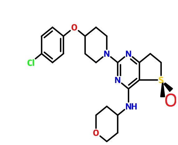
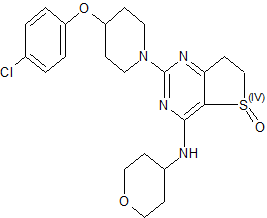
(R)-2-(4-(4-Chlorophenoxy)piperidin-1-yl)-4-((tetrahydro-2H-pyran-4-yl)amino)-6,7-dihydrothieno[3,2-d]pyrimidine 5-Oxide

C22 H27 Cl N4 O3 S, 462.99
 CAS 1910076-27-5
Thieno[3,​2-​d]​pyrimidin-​4-​amine, 2-​[4-​(4-​chlorophenoxy)​-​1-​piperidinyl]​-​6,​7-​dihydro-​N-​(tetrahydro-​2H-​pyran-​4-​yl)​-​, 5-​oxide, (5R)​-

1H NMR (400 MHz, CDCl3) δ 1.49 (dq, J = 4.2, 11.8 Hz, 1H), 1.62 (dq, J = 4.2, 11.8 Hz, 1H), 1.74–1.89 (m, 3H), 1.90–2.02 (m, 3H), 2.96–3.07 (m, 2H), 3.29 (dt, J = 13.6, 8.4 Hz, 1H), 3.44 (ddd, J = 19.2, 11.2, 2.0 Hz, 2H), 3.62 (dt, J = 17.2, 7.8 Hz, 1H), 3.76 (m, 2H), 3.96 (dd, J = 15.6, 12.8 Hz, J = 2H), 4.09–3.99 (m, 3H), 4.51 (m, 1H), 6.21 (br d, J = 6.0 Hz, 1H), 6.86 (d, J = 8.8 Hz, 2H), 7.24 (d, J = 8.8 Hz, 2H);

13C NMR (100 MHz, CDCl3) δ 30.4, 32.5, 32.7, 41.0, 47.2, 49.6, 66.9, 66.9, 72.9, 107.8, 117.5, 125.9, 129.5, 155.8, 158.9, 163.0, 174.6.

The use of phosphodiesterase type 4 (PDE4) inhibitors  for the treatment of COPD (chronic obstructive pulmonary disease) by reducing inflammation and improving lung function is well documented. Given the potential therapeutic benefit offered by these compounds, a number of PDE4-selective inhibitors containing a dihydrothieno[3,2-d]pyrimidine core were identified as preclinical candidates in Boehringer Ingelheim Pharmaceuticals discovery laboratories

While the pathogenesis of chronic obstructive pulmonary disease (COPD) is incompletely understood, chronic inflammation is a major factor. In fact, the inflammatory response is abnormal, with CD8+ T-cells, CD68+ macrophages, and neutrophils predominating in the conducting airways, lung parenchyma, and pulmonary vasculature. Elevated levels of the second messenger cAMP can inhibit some inflammatory processes. Theophylline has long been used in treating asthma; it causes bronchodilation by inhibiting cyclic nucleotide phosphodiesterase (PDE), which inactivates cAMP. By inhibiting PDE, theophylline increases cAMP, inhibiting inflammation and relaxing airway smooth muscle. Rather than one PDE, there are now known to be more than 50, with differing activities, substrate preferences, and tissue distributions. Thus, the possibility exists of selectively inhibiting only the enzyme(s) in the tissue(s) of interest. PDE 4 is the primary cAMP-hydrolyzing enzyme in inflammatory and immune cells (macrophages, eosinophils, neutrophils). Inhibiting PDE 4 in these cells leads to increased cAMP levels, down-regulating the inflammatory response. Because PDE 4 is also expressed in airway smooth muscle and, in vitro, PDE 4 inhibitors relax lung smooth muscle, selective PDE 4 inhibitors are being developed for treating COPD. Clinical studies have been conducted with PDE 4 inhibitors;

Chronic obstructive pulmonary disease (COPD) is a serious and increasing global public health problem; physiologically, it is characterized by progressive, irreversible airflow obstruction and pathologically, by an abnormal airway inflammatory response to noxious particles or gases (MacNee 2005a). The COPD patient suffers a reduction in forced expiratory volume in 1 second (FEV1), a reduction in the ratio of FEV1 to forced vital capacity (FVC), compared with reference values, absolute reductions in expiratory airflow, and little improvement after treatment with an inhaled bronchodilator. Airflow limitation in COPD patients results from mucosal inflammation and edema, bronchoconstriction, increased secretions in the airways, and loss of elastic recoil. Patients with COPD can experience ‘exacerbations,’ involving rapid and prolonged worsening of symptoms (Seneff et al 1995; Connors et al 1996; Dewan et al 2000; Rodriguez-Roisin 2006; Mohan et al 2006). Many are idiopathic, though they often involve bacteria; airway inflammation in exacerbations can be caused or triggered by bacterial antigens (Murphy et al 2000; Blanchard 2002; Murphy 2006;Veeramachaneni and Sethi 2006). Increased IL-6, IL-1β, TNF-α, GRO-α, MCP-1, and IL-8 levels are found in COPD patient sputum; their levels increase further during exacerbations. COPD has many causes and significant differences in prognosis exist, depending on the cause (Barnes 1998; Madison and Irwin 1998).

COPD is already the fourth leading cause of death worldwide, according to the World Health Organization (WHO); the WHO estimates that by the year 2020, COPD will be the third-leading cause of death and the fifth-leading cause of disability worldwide (Murray and Lopez 1997). COPD is the fastest-growing cause of death in developed nations and is responsible for over 2.7 million deaths per year worldwide. In the US, there are currently estimated to be 16 million people with COPD. There are estimated to be up to 20 million sufferers in Japan, which has the world’s highest per capita cigarette consumption and a further 8–12 million in Europe. In 2000, COPD accounted for over 20 million outpatient visits, 3.4 million emergency room visits, 6 million hospitalizations, and 116,500 deaths in the US (National Center for Health Statistics 2002). Factors associated with COPD, including immobility, often lead to secondary health consequences (Polkey and Moxham 2006).

Risk factors for the development of COPD include cigarette smoking, and occupational exposure to dust and chemicals (Senior and Anthonisen 1998; Anthonisen et al 2002; Fabbri and Hurd 2003; Zaher et al 2004). Smoking is the most common cause of COPD and the underlying inflammation typically persists in ex-smokers. Oxidative stress from cigarette smoke is also an issue in COPD (Domej et al 2006). Despite this, relatively few smokers ever develop COPD (Siafakas and Tzortzaki 2002).

While many details of the pathogenesis of COPD remain unclear, chronic inflammation is now recognized as a major factor, predominantly in small airways and lung parenchyma, characterized by increased numbers of macrophages, neutrophils, and T-cells (Barnes 2000; Stockley 2002). As recently as 1995, the American Thoracic Society issued a statement defining COPD without mentioning the underlying inflammation (American Thoracic Society 1995). Since then, the Global Initiative for Chronic Obstructive Lung Disease (GOLD) guidelines have made it clear that chronic inflammation throughout the airways, parenchyma, and pulmonary vasculature plays a central role (Pauwels et al 2001; GOLD 2003). The comparatively recent realization of the role of airway inflammation in COPD has altered thinking with regard to potential therapies (Rogers and Giembycz 1998; Vignola 2004).

Most pharmacological therapies available for COPD, including bronchodilator and anti-inflammatory agents, were first developed for treating asthma. The mainstays of COPD treatment are inhaled corticosteroids (McEvoy and Niewoehner 1998; Borron and deBoisblanc 1998; Pauwels 2002; Gartlehner et al 2006;D’Souza 2006), supplemental oxygen (Petty 1998; Austin and Wood-Baker 2006), inhaled bronchodilators (Costello 1998; Doherty and Briggs 2004), and antibiotics (Taylor 1998), especially in severely affected patients (Anthonisen et al 1987; Saint et al 1995; Adams et al 2001; Miravitlles et al 2002; Donnelly and Rogers 2003; Sin et al 2003; Rabe 2006), though the use of antibiotics remains controversial (Ram et al 2006). Long-acting β2-agonists (LABAs) improve the mucociliary component of COPD. Combination therapy with LABAs and anticholinergic bronchodilators resulted in modest benefits and improved health-related quality of life (Buhl and Farmer 2005; Appleton et al 2006). Treatment with mucolytics reduced exacerbations and the number of days of disability (Poole and Black 2006). The combined use of inhaled corticosteroids and LABAs has been demonstrated to produce sustained improvements in FEV1 and positive effects on quality of life, number of hospitalizations, distance walked, and exacerbations (Mahler et al 2002;Szafranski et al 2003; Sin et al 2004; Miller-Larsson and Selroos 2006; van Schayck and Reid 2006). However, all of these treatments are essentially palliative and do not impact COPD progression (Hay 2000;Gamble et al 2003; Antoniu 2006a).

A further complication in drug development and therapy is that it can be difficult to determine the efficacy of therapy, because COPD has a long preclinical stage, is progressive, and patients generally do not present for treatment until their lung function is already seriously impaired. Moreover, because COPD involves irreversible loss of elasticity, destruction of the alveolar wall, and peribronchial fibrosis, there is often little room for clinical improvement.

Smoking cessation remains the most effective intervention for COPD. Indeed, to date, it is the only intervention shown to stop the decline in lung function, but it does not resolve the underlying inflammation, which persists even in ex-smokers. Smoking cessation is typically best achieved by a multifactor approach, including the use of bupropion, a nicotine replacement product, and behavior modification (Richmond and Zwar 2003).

In COPD, there is an abnormal inflammatory response, characterized by a predominance of CD8+ T-cells, CD68+ macrophages, and neutrophils in the conducting airways, lung parenchyma, and pulmonary vasculature (Soto and Hanania 2005; O’Donnell et al 2006; Wright and Churg 2006). Inflammatory mediators involved in COPD include lipids, inflammatory peptides, reactive oxygen and nitrogen species, chemokines, cytokines, and growth factors. COPD pathology also includes airway remodeling and mucociliary dysfunction (mucus hypersecretion and decreased mucus transport). Corticosteroids reduce the number of mast cells, but CD8+ and CD68+ cells, and neutrophils, are little affected (Jeffery 2005). Inflammation in COPD is not suppressed by corticosteroids, consistent with it being neutrophil-, not eosinophil-mediated. Corticosteroids also do not inhibit the increased concentrations of IL-8 and TNF-α (both neutrophil chemoattractants) found in induced sputum from COPD patients. Neutrophil-derived proteases, including neutrophil elastase and matrix metalloproteinases (MMPs), are involved in the inflammatory process and are responsible for the destruction of elastin fibers in the lung parenchyma (Mercer et al 2005; Gueders et al 2006). MMPs play important roles in the proteolytic degradation of extracellular matrix (ECM), in physiological and pathological processes (Corbel, Belleguic et al 2002). PDE 4 inhibitors can reduce MMP activity and the production of MMPs in human lung fibroblasts stimulated with pro-inflammatory cytokines (Lagente et al 2005). In COPD, abnormal remodeling results in increased deposition of ECM and collagen in lungs, because of an imbalance of MMPs and TIMPs (Jeffery 2001). Fibroblast/myofibroblast proliferation and activation also occur, increasing production of ECM-degrading enzymes (Crouch 1990; Segura-Valdez et al 2000). Additionally, over-expression of cytokines and growth factors stimulates lung fibroblasts to synthesize increased amounts of collagen and MMPs, including MMP-1 (collagenase-1) and MMP-2 and MMP-9 (gelatinases A and B) (Sasaki et al 2000; Zhu et al 2001).

It is now generally accepted that bronchial asthma is also a chronic inflammatory disease (Barnes et al 1988;Barnes 1995). The central role of inflammation of the airways in asthma’s pathogenesis is consistent with the efficacy of corticosteroids in controlling clinical symptoms. Eosinophils are important in initiating and continuing the inflammatory state (Holgate et al 1987; Bruijnzeel 1989; Underwood et al 1994; Teixeira et al 1997), while other inflammatory cells, including lymphocytes, also infiltrate the airways (Holgate et al 1987;Teixeira et al 1997). The familiar acute symptoms of asthma are the result of airway smooth muscle contraction. While recognition of the key role of inflammation has led to an emphasis on anti-inflammatory therapy in asthma, a significant minority of patients remains poorly controlled and some exhibit accelerated declines in lung function, consistent with airway remodeling (Martin and Reid 2006). Reversal or prevention of structural changes in remodeling may require additional therapy (Burgess et al 2006).

There is currently no cure for asthma; treatment depends primarily on inhaled glucocorticoids to reduce inflammation (Taylor 1998; Petty 1998), and inhaled bronchodilators to reduce symptoms (Torphy 1994;Costello 1998; Georgitis 1999; DeKorte 2003). Such treatments, however, do not address disease progression.

COPD and asthma are both characterized by airflow obstruction, but they are distinct in terms of risk factors and clinical presentation. While both involve chronic inflammation and cellular infiltration and activation, different cell types are implicated and there are differences in the inflammatory states (Giembycz 2000;Fabbri and Hurd 2003; Barnes 2006). In COPD, neutrophil infiltration into the airways and their activation appear to be key (Stockley 2002); in asthma, the inflammatory response involves airway infiltration by activated eosinophils and lymphocytes, and T-cell activation of the allergic response (Holgate et al 1987;Saetta et al 1998; Barnes 2006). While macrophages are present in both conditions, the major controller cells are CD8+ T-cells in COPD (O’Shaughnessy et al 1997; Saetta et al 1998) and CD4+ T-cells in asthma. IL-1, IL-8, and TNF-α are the key cytokines in COPD, while in asthma, IL-4, IL-5, and IL-13 are more important. There are differences in histopathological features of lung biopsies between COPD patients and asthmatics; COPD patients have many fewer eosinophils in lung tissue than asthmatics.

While the early phases of COPD and asthma are distinguishable, there are common features, including airway hyper-responsiveness and mucus hypersecretion. MUC5AC is a major mucin gene expressed in the airways; its expression is increased in COPD and asthmatic patients. At least in vitro, epidermal growth factor stimulates MUC5AC mRNA and protein expression; this can be reversed by PDE 4 inhibitors, which may contribute to their clinical efficacy in COPD and asthma (Mata et al 2005). Similar structural and fibrotic changes make COPD and asthma much less distinguishable in extreme cases; the chronic phases of both involve inflammatory responses, alveolar detachment, mucus hypersecretion, and subepithelial fibrosis. The two conditions have been linked epidemiologically; adults with asthma are up to 12 times more likely to develop COPD over time than those without (Guerra 2005).


PAPER


Abstract Image

A practical, safe, and efficient process for the synthesis of PDE4 (phosphodiesterase type 4) inhibitors represented by 1 and 2 was developed and demonstrated on a multi-kilogram scale. Key aspects of the process include the regioselective synthesis of dihydrothieno[3,2-d]pyrimidine-2,4-diol 9 and the asymmetric sulfur oxidation of intermediate 11.

Development of a Practical Process for the Synthesis of PDE4 Inhibitors

Chemical Development US, Boehringer Ingelheim Pharmaceuticals, Inc., 900 Ridgebury Road, P.O. Box 368, Ridgefield, Connecticut 06877-0368, United States
Org. Process Res. Dev., Article ASAP
DOI: 10.1021/acs.oprd.6b00104


PDE 4 in COPD

With regard to COPD, PDE 4 is the primary cAMP-hydrolyzing enzyme in inflammatory and immune cells, especially macrophages, eosinophils, and neutrophils, all of which are found in the lungs of COPD and asthma patients (Torphy et al 1992; Karlsson and Aldous 1997; De Brito et al 1997; Wang et al 1999;Torphy and Page 2000). Inhibition of PDE 4 leads to elevated cAMP levels in these cells, down-regulating the inflammatory response (Dyke and Montana 2002).

PDE 4 has also attracted much attention because it is expressed in airway smooth muscle (Ashton et al 1994;Undem et al 1994; Nicholson et al 1995; Kerstjens and Timens 2003; Mehats et al 2003; Lipworth 2005; Fan Chung 2006). In vitro, PDE 4 inhibitors relax lung smooth muscle (Undem et al 1994; Dent and Giembycz 1995). In COPD and asthma, a selective PDE 4 inhibitor with combined bronchodilatory and anti-inflammatory properties would seem desirable (Nicholson and Shahid 1994; Lombardo 1995; Palfreyman 1995; Cavalia and Frith 1995; Palfreyman and Souness 1996; Karlsson and Aldous 1997; Compton et al 2001; Giembycz 2002; Jacob et al 2002; Soto and Hanania 2005).

PDE 4 inhibitors in COPD

So, because PDE 4 inhibitors suppress inflammatory functions in several cell types involved in COPD and asthma (Huang and Mancini 2006) and because, at least in vitro, PDE 4 inhibitors relax lung smooth muscle, selective PDE 4 inhibitors, originally intended for use in treating depression (Renau 2004), have been developed for the treatment of COPD and asthma (Torphy et al 1999; Spina 2000; Huang et al 2001; Spina 2004; Giembycz 2005a, 2005b; Lagente et al 2005; Boswell-Smith, Spina et al 2006). PDE 4 enzymes are strongly inhibited by the antidepressant drug rolipram (Pinto et al 1993), which decreases the influx of inflammatory cells at sites of inflammation (Lagente et al 1994; Lagente et al 1995; Alves et al 1996). PDE 4 inhibitors down-regulate cytokine production in inflammatory cells, in vivo and in vitro (Undem et al 1994;Dent and Giembycz 1995). TNF-α is an important inflammatory cytokine in COPD; its release is reduced by PDE 4 inhibitors (Souness et al 1996; Chambers et al 1997; Griswold et al 1998; Gonçalves de Moraes et al 1998; Corbel, Belleguic et al 2002). Some PDE 4 inhibitors, including cilomilast and AWD 12-281, can inhibit neutrophil degranulation, a property not shared by theophylline (Ezeamuzie 2001; Jones et al 2005). PDE 4 inhibitors reduce overproduction of other pro-inflammatory mediators, including arachidonic acid and leukotrienes (Torphy 1998). PDE 4 inhibitors also inhibit cellular trafficking and microvascular leakage, production of reactive oxygen species, and cell adhesion molecule expression in vitro and in vivo (Sanz et al 2005). PDE 4 inhibitors, including cilomilast and CI-1044, inhibit LPS-stimulated TNF-α production in whole blood from COPD patients (Burnouf et al 2000; Ouagued et al 2005).

There are now thought to be at least four PDE 4s, A, B, C, and D, derived from four genes (Lobbam et al 1994; Muller et al 1996; Torphy 1998; Conti and Jin 1999; Matsumoto et al 2003). Alternative splicing and alternative promoters add further complexity (Manganiello et al 1995; Horton et al 1995; Torphy 1998). Indeed, the four genes encode more than 16 PDE 4 isoforms, which can be divided into short (∼65–75 kDa) and long forms (∼80–130 kDa); the difference between the short and long forms lies in the N-terminal region (Bolger et al 1997; Huston et al 2006). PDE 4 isoforms are regulated by extracellular signal-related protein kinase (ERK), which can phosphorylate PDE 4 (Houslay and Adams 2003).

The four PDE 4 genes are differentially expressed in various tissues (Silver et al 1988; Lobbam et al 1994;Manganiello et al 1995; Horton et al 1995; Muller et al 1996; Torphy 1998). PDE 4A is expressed in many tissues, but not in neutrophils (Wang et al 1999). PDE 4B is also widely expressed and is the predominant PDE 4 subtype in monocytes and neutrophils (Wang et al 1999), but is not found in cortex or epithelial cells (Jin et al 1998). Upregulation of the PDE 4B enzyme in response to pro-inflammatory agents suggest that it has a role in inflammatory processes (Manning et al 1999). PDE 4C is expressed in lung and testis, but not in circulating inflammatory cells, cortex, or hippocampus (Obernolte et al 1997; Manning et al 1999; Martin-Chouly et al 2004). PDE 4D is highly expressed in lung, cortex, cerebellum, and T-cells (Erdogan and Houslay 1997; Jin et al 1998). PDE 4D also plays an important role in airway smooth muscle contraction (Mehats et al 2003).

A major issue with early PDE 4 inhibitors was their side effect profile; the signature side effects are largely gastrointestinal (nausea, vomiting, increased gastric acid secretion) and limited the therapeutic use of PDE 4 inhibitors (Dyke and Montana 2002). The second generation of more selective inhibitors, such as cilomilast and roflumilast, have improved side effect profiles and have shown clinical efficacy in COPD and asthma (Barnette 1999; Spina 2000; Lagente et al 2005). However, even cilomilast and roflumilast, the most advanced clinical candidates, discussed below, cause some degree of emesis (Spina 2003).

It is now thought that the desirable anti-inflammatory properties and unwanted side effects of nausea and emesis are associated with distinct biochemical activities (Torphy et al 1992; Jacobitz et al 1996; Barnette et al 1996; Souness et al 1997; Souness and Rao 1997). Specifically, the side effects are believed to be associated with the so-called ‘high-affinity rolipram binding site’ (HARBS) (Barnette et al 1995; Muller et al 1996; Jacobitz et al 1996; Kelly et al 1996; Torphy 1998) and/or inhibition of the form of PDE 4 found in the CNS (Barnette et al 1996). The exact nature of HARBS remains unclear, although it has been described as a conformer of PDE 4 (Souness and Rao 1997; Barnette et al 1998). Using mice deficient in PDE 4B or PDE 4D, it appears that emesis is the result of selective inhibition of PDE 4D (Robichaud et al 2002; Lipworth 2005), which is unfortunate, because the most clinically advanced PDE 4 inhibitors are selective for PDE 4D. Also, from animal studies, it appears that the nausea and vomiting are produced via the CNS, though there may also be direct effects on the gastrointestinal system (Barnette 1999).

While beyond the scope of this review, it has been proposed that PDE 4 inhibitors may be useful in treating inflammatory bowel disease (Banner and Trevethick 2004), cystic fibrosis (Liu et al 2005), pulmonary arterial hypertension (Growcott et al 2006), myeloid and lymphoid malignancies (Lerner and Epstein 2006), Alzheimer’s disease (Ghavami et al 2006), rheumatoid arthritis and multiple sclerosis (Dyke and Montana 2002), infection-induced preterm labor (Oger et al 2004), depression (Wong et al 2006), and allergic disease (Crocker and Townley 1999). Varying degrees of in vitro, in vivo, and clinical data exist to support these claims.

So, after that theoretical buildup, we reach the proof of the pudding; clinical studies have been conducted with PDE 4 inhibitors. A potent, but not-very-selective, PDE 4 inhibitor is approved in Japan and is used clinically, including for treating asthma. Another is awaiting approval in the US. One is in advanced clinical development and others are at earlier stages.

REF

Pouzet, P.; Hoenke, C.; Martyres, D.; Nickolaus, P.; Jung, B.; Hamman, H. Dihydrothienopyrimidines for the treatment of inflammatory diseases. PatentWO 2006111549 A1, October 26, 2006.

Ohnacker, G.; Woitun, E. Novel dihydrothieno[3, 2-d]pyrimidines. U.S. Patent US 3,318,881, May 9, 1967.

/////PDE4 Inhibitors, Boehringer Ingelheim Pharmaceuticals, BI ?, PRECLINICAL, 1910076-27-5

Clc1ccc(cc1)OC2CCN(CC2)c4nc(NC3CCOCC3)c5c(n4)CCS5=O

Share

MK 8718

 Uncategorized  Comments Off on MK 8718
May 172016
 

img

Figure imgf000105_0002

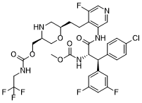
MK 8718

Cas 1582729-24-5 (free base); 1582732-29-3 (HCl).
MF: C30H30ClF6N5O4
MW: 673.1891

INNOVATOR Merck Sharp & Dohme Corp., Merck Canada Inc.

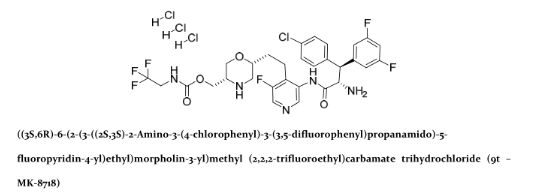
((3S,6R)-6-(2-(3-((2S,3S)-2-amino-3-(4-chlorophenyl)-3-(3,5-difluorophenyl)propanamido)-5-fluoropyridin-4-yl)ethyl)morpholin-3-yl)methyl (2,2,2-trifluoroethyl)carbamate

MK-8718 is a potent, selective and orally bioavailable HIV protease inhibitor with a favorable pharmacokinetic profile with potential for further development.

A retrovirus designated human immunodeficiency virus (HIV), particularly the strains known as HIV type-1 (HIV-1) virus and type-2 (HIV-2) virus, is the etiological agent of acquired immunodeficiency syndrome (AIDS), a disease characterized by the destruction of the immune system, particularly of CD4 T-cells, with attendant susceptibility to opportunistic infections, and its precursor AIDS-related complex (“ARC”), a syndrome characterized by symptoms such as persistent generalized lymphadenopathy, fever and weight loss. This virus was previously known as LAV, HTLV-III, or ARV. A common feature of retrovirus replication is the extensive post-translational processing of precursor polyproteins by a virally encoded protease to generate mature viral proteins required for virus assembly and function. Inhibition of this processing prevents the production of normally infectious virus. For example, Kohl et al., Proc. Nat’l Acad. Sci. 1988, 85: 4686, demonstrated that genetic inactivation of the HIV encoded protease resulted in the production of immature, non-infectious virus particles. These results indicated that inhibition of the HIV protease represents a viable method for the treatment of AIDS and the prevention or treatment of infection by HIV.

Nucleotide sequencing of HIV shows the presence of a pol gene in one open reading frame [Ratner et al, Nature 1985, 313: 277]. Amino acid sequence homology provides evidence that the pol sequence encodes reverse transcriptase, an endonuclease, HIV protease and gag, which encodes the core proteins of the virion (Toh et al, EMBO J. 1985, 4: 1267; Power et al, Science 1986, 231 : 1567; Pearl et al, Nature 1987, 329: 351].

Several HIV protease inhibitors are presently approved for clinical use in the treatment of AIDS and HIV infection, including indinavir (see US 5413999), amprenavir (US5585397), saquinavir (US 5196438), ritonavir (US 5484801) and nelfmavir (US 5484926). Each of these protease inhibitors is a peptide-derived peptidomimetic, competitive inhibitor of the viral protease which prevents cleavage of the HIV gag-pol polyprotein precursor. Tipranavir (US 5852195) is a non-peptide peptidomimetic protease inhibitors also approved for use in treating HIV infection. The protease inhibitors are administered in combination with at least one and typically at least two other HIV antiviral agents, particularly nucleoside reverse transcriptase inhibitors such as zidovudine (AZT) and lamivudine (3TC) and/or non-nucleoside reverse transcriptase inhibitors such as efavirenz and nevirapine. Indinavir, for example, has been found to be highly effective in reducing HIV viral loads and increasing CD4 cell counts in HIV-infected patients, when used in combination with nucleoside reverse transcriptase inhibitors. See, for example, Hammer et al, New England J. Med. 1997, 337: 725-733 and Gulick et al, New England J. Med. 1997, 337: 734-739.

The established therapies employing a protease inhibitor are not suitable for use in all HIV-infected subjects. Some subjects, for example, cannot tolerate these therapies due to adverse effects. Many HIV-infected subjects often develop resistance to particular protease inhibitors. Furthermore, the currently available protease inhibitors are rapidly metabolized and cleared from the bloodstream, requiring frequent dosing and use of a boosting agent.

Accordingly, there is a continuing need for new compounds which are capable of inhibiting HIV protease and suitable for use in the treatment or prophylaxis of infection by HIV and/or for the treatment or prophylaxis or delay in the onset or progression of AIDS.

str1

str1

PATENT

https://www.google.co.in/patents/WO2014043019A1?cl=en

INTERMEDIATE 1

Synthesis of morpholine intermediate (tert-butyl ( ^S^-S-d tert- butyl(dimethyl)silylloxy|methyl)-2-(hydroxymethyl)morpholine-4-carboxylate)

Scheme 1

EXAMPLE 97

( S)- -(4-Chlorophenyl)-3,5-difiuoro-N-(5-fiuoro-4-{2-[(2R,5S)-5-({[(2,2,2- trifluoroethyl)carbamoyl]oxy}methyl)morpholin-2-yl]ethyl}pyridin-3-yl)-L-phenylalaninamide

Step 1. (2S,3S)-2-Azido-3-(4-chlorophenyl)-3-(3,5-difluorophenyl)propanoic acid

The title compound was prepared from 4-chlorocinnamic acid and 3,5- difluorophenylmagnesium bromide using the procedures given in steps 1-4 of Example 92.

Step 2. (2R,5S)-tert-butyl 2-(2-(3-((2S,3S)-2-azido-3-(4-chlorophenyl)-3-(3,5- difluorophenyl)propanamido)-5-fluoropyridin-4-yl)ethyl)-5-((((2,2,2- trifluoroethyl)carbamoyl)oxy)methyl)morpholine-4-carboxylate

The product from step 1 (105 mg, 0.31 mmol) and the product from step 4 of Example 89 (150 mg, 0.31 mmol) were dissolved in pyridine (1 mL) and the stirred solution was cooled to -10 °C in an ice/acetone bath. To the cold solution was added POCI3 dropwise (0.035 mL, 0.38 mmol). The mixture was stirred at -10 °C for 30 min. The reaction was quenched by the addition of saturated aqueous NaHC03 solution (1 mL) and the mixture was allowed to warm to ambient temperature. The mixture was diluted with water (10 mL) and extracted with dichloromethane (3 x 10 mL). The combined dichloromethane phases were dried (Na2S04), filtered, and the filtrate solvents were removed in vacuo. The residue was purified on a 12 g silica gel column using a gradient elution of 0-70% EtOAc:hexanes. Fractions containing product were combined and the solvents were removed in vacuo to give the title compound as a gum. (M+H)+ = 800.6.

Step 3. (2R,5S)-tert-butyl 2-(2-(3-((2S,3S)-2-amino-3-(4-chlorophenyl)-3-(3,5- difluorophenyl)propanamido)-5-fluoropyridin-4-yl)ethyl)-5-((((2,2,2- trifluoroethyl)carbamoyl)oxy)methyl)morpholine-4-carboxylate

The product from step 2 (150 mg, 0.19 mmol) and triphenylphosphine (74 mg, 0.28 mmol) were dissolved in THF (4 mL) and to the solution was added water (1 mL). The mixture was heated to reflux under a nitrogen atmosphere for 12 h. The mixture was cooled to ambient temperature and the solvents were removed in vacuo. The residue was purified on a 12 g silica gel column eluting with a gradient of 0-10% methanol: chloroform. Fractions containing product were combined and the solvents were removed in vacuo to give the title compound as a gum. (M+H)+ = 774.7. Step 4. ( S)- -(4-Chlorophenyl)-3,5-difluoro-N-(5-fluoro-4-{2-[(2R,5S)-5-({[(2,2,2- trifluoroethyl)carbamoyl]oxy}methyl)morpholin-2-yl]ethyl}pyridin-3-yl)-L-phenylala

The product from step 3 (60 mg, 0.078 mmol) was dissolved in a solution of 4M HCl in dioxane (1 mL, 4 mmol) and the solution was stirred at ambient temperature for 1 h. The solvent was removed under reduced pressure and the residue was dried in vacuo for 12 h to give an HCl salt of the title compound as a solid. LCMS: RT = 0.95 min (2 min gradient), MS (ES) m/z = 674.6 (M+H)+.


PAPER

Abstract Image

A novel HIV protease inhibitor was designed using a morpholine core as the aspartate binding group. Analysis of the crystal structure of the initial lead bound to HIV protease enabled optimization of enzyme potency and antiviral activity. This afforded a series of potent orally bioavailable inhibitors of which MK-8718 was identified as a compound with a favorable overall profile.

Discovery of MK-8718, an HIV Protease Inhibitor Containing a Novel Morpholine Aspartate Binding Group

Merck Research Laboratories, 770 Sumneytown Pike, PO Box 4, West Point, Pennsylvania 19486, United States
Merck Frosst Centre for Therapeutic Research, 16711 TransCanada Highway, Kirkland, Quebec H9H 3L1, Canada
§Albany Molecular Research Singapore Research Center, 61 Science Park Road #05-01, The Galen Singapore Science Park II, Singapore 117525
ACS Med. Chem. Lett., Article ASAP
DOI: 10.1021/acsmedchemlett.6b00135
*E-mail: christopher_bungard@merck.com. Phone: 215-652-5002.

References

Discovery of MK-8718, an HIV Protease Inhibitor Containing a Novel Morpholine Aspartate Binding Group
Christopher J. Bungard*†, Peter D. Williams†, Jeanine E. Ballard†, David J. Bennett†, Christian Beaulieu‡, Carolyn Bahnck-Teets†, Steve S. Carroll†, Ronald K. Chang†, David C. Dubost†, John F. Fay†, Tracy L. Diamond†, Thomas J. Greshock†, Li Hao§, M. Katharine Holloway†, Peter J. Felock, Jennifer J. Gesell†, Hua-Poo Su†, Jesse J. Manikowski†, Daniel J. McKay‡, Mike Miller†, Xu Min†, Carmela Molinaro†, Oscar M. Moradei‡, Philippe G. Nantermet†, Christian Nadeau‡, Rosa I. Sanchez†, Tummanapalli Satyanarayana§, William D. Shipe†, Sanjay K. Singh§, Vouy Linh Truong‡, Sivalenka Vijayasaradhi§, Catherine M. Wiscount†, Joseph P. Vacca‡, Sheldon N. Crane‡, and John A. McCauley†
† Merck Research Laboratories, 770 Sumneytown Pike, PO Box 4, West Point, Pennsylvania 19486, United States
‡ Merck Frosst Centre for Therapeutic Research, 16711 TransCanada Highway, Kirkland, Quebec H9H 3L1, Canada
§ Albany Molecular Research Singapore Research Center, 61 Science Park Road #05-01, The Galen Singapore Science Park II, Singapore 117525
ACS Med. Chem. Lett., Article ASAP
DOI: 10.1021/acsmedchemlett.6b00135
Publication Date (Web): May 09, 2016

////MK-8718, HIV, protease, inhibitor

Supporting Info

O=C(OC[C@H]1NC[C@@H](CCC(C(F)=CN=C2)=C2NC([C@@H](N)[C@@H](C3=CC=C(Cl)C=C3)C4=CC(F)=CC(F)=C4)=O)OC1)NCC(F)(F)F

Share

MK-7145

 Uncategorized  Comments Off on MK-7145
May 172016
 

2D chemical structure of 1255204-84-2

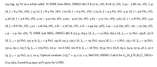
MK-7145,

cas  1255204-84-2

1(3H)-Isobenzofuranone, 5,5′-(1,4-piperazinediylbis((1R)-1-hydroxy-2,1-ethanediyl))bis(4-methyl-

MF C26 H30 N2 O6, Molecular Weight 466.53
1(3H)​-​Isobenzofuranone, 5,​5′-​[1,​4-​piperazinediylbis[(1​R)​-​1-​hydroxy-​2,​1-​ethanediyl]​]​bis[4-​methyl-

The Renal Outer Medullary Potassium (ROMK) channel (KM .1 ) (see e.g., Ho,K., et al., Cloning and expression of an inwardly rectifying ATP -regulated potassium channel, Nature, 1993, 362(6415): p. 31-8.1, 2; and Shuck, M.E., et al., Cloning and characterization of multiple forms of the human kidney ROM-K potassium channel, J Biol Chem, 1994, 269(39): p. 24261-70) is a member of the inward rectifier family of potassium channels expressed in two regions of the kidney: thick ascending loop of Henle (TALH) and cortical collecting duct (CCD) (see Hebert, S. C, et al., Molecular diversity and regulation of renal potassium channels, Physiol Rev, 2005, 85(1): p. 319-713). At the TALH, ROMK participates in potassium recycling across the luminal membrane which is critical for the function of the Na+/K+/2CF co-transporter, the rate-determining step for salt reuptake in this part of the nephron. At the CCD, ROMK provides a pathway for potassium secretion that is tightly coupled to sodium uptake through the amiloride-sensitive sodium channel (see Reinalter, S. C, et al., Pharmacotyping of hypokalemic salt-losing tubular disorders, Acta. Physiol Scand, 2004, 181(4): p. 513-21 ; and Wang, W., Renal potassium channels: recent developments, Curr Opin Nephrol Hypertens, 2004, 13(5): p. 549-55). Selective inhibitors of the ROMK channel (also referred to herein as inhibitors of ROMK or ROMK inhibitors) are predicted to represent novel diuretics for the treatment of hypertension and other conditions where treatment with a diuretic would be beneficial with potentially reduced liabilities (i.e., hypo- or hyperkalemia, new onset of diabetes, dyslipidemia) over the currently used clinical agents (see Lifton, R.P., A.G. Gharavi, and D.S. Geller, Molecular mechanisms of human hypertension, Cell, 2001, 104(4): p. 545-56). Human genetics (Ji, W., et al., Rare independent mutations in renal salt handling genes contribute to blood pressure variation, Nat Genet, 2008, 40(5): p. 592-9; and Tobin, M.D., et al., Common variants in genes underlying monogenic hypertension and hypotension and blood pressure in the general population, Hypertension, 2008, 51(6): p. 1658-64) and genetic ablation of ROMK in rodents (see Lorenz, J.N., et al., Impaired renal NaCl absorption in mice lacking the ROMK potassium channel, a model for type II Bartter’s syndrome, J Biol Chem, 2002, 277(40): p. 37871-80 and Lu, M., et al.s Absence of small conductance K+ channel (SK) activity in apical membranes of thick ascending limb and cortical collecting duct in ROMK (Banter’s) knockout mice, J Biol Chem, 2002, 277(40): p. 37881-7) support these expectations. To our knowledge, the first small molecule selective inhibitors of ROMK were reported from work done at Vanderbilt University as described in Lewis, L.M., et al., High-Throughput Screening Reveals a Small-Molecule Inhibitor of the Renal Outer Medullary Potassium Channel and KirJ.l, MoI Pharmacol, 2009, 76(5): p. 1094-1103.

PATENT

WO 2010129379

http://www.google.com/patents/WO2010129379A1?cl=ko

SCHEME 1


SCHEME 2


SCHEME 3


SCHEME 5


SCHEME 6

SCHEME 7


SCHEME 8


14 15

The preparation of compounds 16 can be achieved following the sequence detailed in Scheme 9. Treating epoxide 2-1 with commercially available 1-Boc piperazine at elevated temperatures gives rise to alcohol 2-2 (Nomura, Y. et al. Chemical & Pharmaceutical Bulletin, 1995, 43(2), 241-6). The hydroxyl group of 2-2 can be converted to the fluoride by treatment of such fluorinating reagent as DAST (Hudlicky, M. Organic Reactions, 1988, 35). Removal of the Boc group of 3-1 under acidic conditions such as TFA gives rise to piperazine 3-2. Piperazine 3-2 can be washed with an aqueous base solution followed by extraction with organic solvents to generate the free base form. The free base of 3-2 can be coupled to epoxide 5-1 at elevated temperatures to afford compound 16. The Ar-CHF- and Ar’-CHOH- groups in 16 represent examples of either Z1 or Z2.

SCHEME 9


16 General Procedures.

INTERMEDIATE (Ry-H (free base)

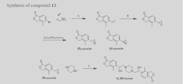
5-\(lR)-l -hγdroxγ-2-piperazio- 1 -ylethyl] -4-methyl-2-benzofuran- 1 f 3/f)-one To a 20 mL microwave tube charged with 4-methyl-5-[(2jS)-oxiran-2-yl]-2-benzofuran-l(3H)-one (1020 mg, 5.40 mmol) and a stir bar was added 1-Boc Piperazine (800mg, 4.3 mmol) and EtOH (15 mL). The tube was sealed and heated in a microwave apparatus to 150 0C for 1 hour. The crude product was adsorbed onto silica gel, and purified by flash chromatography (Hexanes-EtOAc with 10% EtOH: 0 – 100% gradient), and solvent removed to afford terl-butyl~4-[(2R-2-hydroxy-2-(4-methyl-l -oxo-1 ,3-dihydro-2-bers2θfuran-5-yl) ethyl}piperazine-l-carboxylate. LCMS M+l (calc. 377.20, found 377.13). This product was treated with neat TFA for 15 minutes to remove the Boc group. After removal of TFA under reduced pressure, the residue was taken into aq NaHCO3, and back-extracted with CHCl3-IPA (3:1). The organic layers were combined, dried over sodium sulfate, and concentrated to afford 5 – [( 1 R)- 1 -hydroxy-2-piperazin- 1 -ylethyl] -4-methyl-2-benzofuran- 1 (3H)-one. 1H NMR (OMSO-d6, 500 MHz) δ 7.68 (d, J= 8.0 Hz, IH), 7.65 (d, J= 8.0 Hz, IH)5 5.38, 5.35 (AB system, J- 15.4, J= 16.7, 2H), 5.06 (dd5 J- 3.9 Hz, J= 3.7 Hz, IH), 3.76 (m, IH)5 2.72 (m, 4H), 2.42 (m, 4H), 2.34 (d, J= 3.8 Hz5 IH), 2.32 (d, J= 3.8 Hz, IH), 2.24 (s, 3H); LC/MS: (IE, m/z) [M +I]+ = 277.03.

EXAMPLE 2A

5, 5 ‘-{ piperazine- 1 ,4-diylbis[( 1 R)- 1 -hydroxy ethane-2 , 1 -diyl] } bis(4-methyl-2-benzofuran- 1 (3H)-one)

Method 1: To a 20 mL microwave tube charged with 4-methyl-5-[(2i?)-oxiran-2-yl]-2-benzofuran-l(3H)-one (972 mg, 5.11 mmol) and piperazine (200 mg, 2.3 mmol) was added a stir bar and EtOH (16 mL). The tube was sealed and heated in a microwave apparatus to 150 0C for 90 minutes. The crude product was adsorbed onto silica gel, and purified by flash chromatography (MeOΗ-DCM 0 ~ 7% gradient). After removal of solvents, 5»5′-{piperazine-1 ,4-diyIbi s [( 1 R)- 1 -hydroxyethane-2, 1 -diyl] } bis(4-methyl-2-benzofuran- 1 (3 H)-one) was collected. 1H-NMR (500 MHz9 CDCl3) δ ppm 7.80 (s, 4H), 5.25 (s, 4H), 5.11 (d, J= 10.5 Hz5 2H), 4.00 (broad, 2H), 2.90 (broad, 4H)3 2.69-2.50 (m, 6H), 2.44 (t, J= 11 Hz, 2H), 2.29 (s, 6H); LCMS M+l (calc. 467, found 467).

Method 2: Piperazine (4.51 g, 52.4 mmol) and 4-methyl-5-[(2Λ)-oxiran-2-yl]-2-benzofuran-1 (3//)-one (20.0 g, 105 mmol) were charged to a 3-neck 500-mL roundbottom flask, equipped with a reflux condensor, under nitrogen. Toluene (80.0 mL, 751 mmol) and N,N-dimethylacetamide (80 mL, 854 mmol) were added to provide a suspension. The reaction mixture was warmed to 110 0C, becoming homogeneous at 25 0C. After stirring for 4.5 h at 110 0C, the temperature was increased to 115 °C to drive the reaction forward. After stirring for 48 h, the reaction mixture was cooled to RT. On cooling, crystallization occurred. Water was added via addition funnel (45 mL), generating a thick slurry. The suspension was filtered and the solids were washed with 4:1 water :DMA (60 mL), followed by water (2 x 35 mL). The solid was dried on the funnel under vacuum with a nitrogen sweep to constant mass. 5,5′-{Piperazine-l,4-diylbis[(li?)-l-hydroxyethane-2,l-diyl]}bis(4-methyl-2-beiizofurari-l(3H)-one) was isolated. 1H-NMR (500 MHz, CDCl3) δ ppm 7.80 (s, 4H), 5.25 (s, 4H), 5.11 (d, J- 11 Hz, 2H), 4.30-3.51 (broad, 2H), 2.90 (broad, 4H), 2.69-2.50 (m, 6H), 2.44 (t, J- 11 Hz, 2H), 2.30 (s, 6H).

Compounds of the present invention are amines and can therefore be converted to a variety of salts by treatment with any of a number of acids. For example, the compound of Example 2A can be converted to several different salt forms as shown in the following representative examples. These are selected examples and are not meant to be an exhaustive list; numerous additional salts can be prepared in a similar fashion using a variety of acids. EXAMPLE 2A-1 (di-HCl salt): 5,5t-{piperazme-l,4-diylbis[(17?)-l-hydroxyethane-2,l- diyl] } bis(4-methyl-2-benzofuran- 1 (3H)-one) dihydrochloride To a 250 mL pear shape flask charged with the free base (1.2 g, 2.6 mmol) and a stir bar was added DCM. The solution was stirred until all solids were gone. To this solution was added 4N HCl in dioxane (2.6 mL, 4.0 eq), and the mixture was allowed to stir for another 15 minutes. The solvent was removed on a rotary evaporator, and the product was left dry on a high vacuum pump until there was no weight change. The product was determined to be 5, 5 ‘-{piperazine- 1,4-diylbis [( 1 R)- 1 ~hydroxyethane-2, 1 -diyl] } bis(4-methyl-2-benzofuran- 1 (3i?)-one) dihydrochloride. EXAMPLE 2A-2 (HCl salt): 5,5’-{piperazine-l,4-diylbis[(l^)-l-hydroxyethane-2,l- diyl] } bis(4-methyl-2-benzofuran- 1 QHVone) hvdrochl oride

To a 20 dram vial charged with the free base (160 mg, 0.34 mmol) and a stir bar was added 0.1 M HCl in IPA. The solution was allowed to stir at RT for 30 minutes, and then heated to 400C for 1 hour. The solvent was removed under vacuum, and the resulting product was left on a high vacuum pump for 16 hours. The product corresponded to 5,5′-{piperazine-l,4-diylbis[(li?)-l-hydroxyethane~2, 1 -diyl] } bis(4-methyl-2-benzofuran- 1 (3 H)-one) hydrochloride.

EXAMPLE 2A-3 (mono-hydrate of the di-HCl salt): 5, 5′- {piperazine- l,4-diylbis[( Ii?)- 1-hydroxyethane-2,l-diyl] Ibis^-niethyl-g-benzofuran-lfS/^-one) dihydrochloride hydrate To a flask charged with the free base (1.0 g, 2.1 rnmol) and a stir bar was added 1 N HCl (50 mL). The mixture was allowed to stir until all solids dissolved. The solvent was removed on a rotary evaporator, and the resulting product was left on a high vacuum pump for 16 hours. The product was determined to be 5,5′-{piperazine-l ,4-diylbis[(li?)-l-hydroxyethane-2,l-diyl]}bis(4-methyl-2-benzofuran-l(3H)-one) dihydrochloride hydrate.

EXAMPLE 2A-4 (H2SO4 salt): 5.5′-{piperaziiie-l>4-diylbis[(lJΪ)-l-hydioxyethane-2,l- diyl] }bis(4-methyl-2-benzofuran-l(3/f)-one) sulfate (salt) To a 100 mL flask charged with a solution of the free base (154 mg, 0.330 mmol) in DMF : MeOH (3 : 1) (20 mL) and a stir bar was added 0.1 M H2SO4 (3.3 mL). The solution was allowed to stir at RT for 30 minutes, and then heated to 40 0C for 2 hours. A lot of solids formed during that time. The solvent was removed under vacuum, and the white solids were left on high vacuum for 16 hours to afford 5)5l-{piperazine-l,4-diylbis[(lJ?)~l-hydroxyethane-2,l-diyl] }bis(4-methyl-2-benzofuran-l(3H)-one) sulfate (salt).

Paper

Abstract Image

ROMK, the renal outer medullary potassium channel, is involved in potassium recycling at the thick ascending loop of Henle and potassium secretion at the cortical collecting duct in the kidney nephron. Because of this dual site of action, selective inhibitors of ROMK are expected to represent a new class of diuretics/natriuretics with superior efficacy and reduced urinary loss of potassium compared to standard-of-care loop and thiazide diuretics. Following our earlier work, this communication will detail subsequent medicinal chemistry endeavors to further improve lead selectivity against the hERG channel and preclinical pharmacokinetic properties. Pharmacological assessment of highlighted inhibitors will be described, including pharmacodynamic studies in both an acute rat diuresis/natriuresis model and a subchronic blood pressure model in spontaneous hypertensive rats. These proof-of-biology studies established for the first time that the human and rodent genetics accurately predict the in vivo pharmacology of ROMK inhibitors and supported identification of the first small molecule ROMK inhibitor clinical candidate, MK-7145.

Discovery of MK-7145, an Oral Small Molecule ROMK Inhibitor for the Treatment of Hypertension and Heart Failure

Departments of Discovery Chemistry, Ion Channels, §In Vivo Pharmacology, Cardiorenal, and Pharmacokinetics, Pharmacodynamics and Drug Metabolism, Merck Research Laboratories, Kenilworth, New Jersey 07033, United States
ACS Med. Chem. Lett., Article ASAP
DOI: 10.1021/acsmedchemlett.6b00122
*Tel: 908-740 4932. E-mail: haifeng_tang@merck.com.
str1

////////

Cc1c(ccc2c1COC2=O)[C@H](CN3CCN(CC3)C[C@@H](c4ccc5c(c4C)COC5=O)O)O

Share

ICH Q3D Implementation Working Group (IWG)—Training Modules

 regulatory, Uncategorized  Comments Off on ICH Q3D Implementation Working Group (IWG)—Training Modules
May 172016
 

ICH Q3D Implementation Working Group (IWG)—Training Modules


ICH Q3D is a complex guideline. The overall requirement in terms of control is clear—there are defined limits for some 24 elements, and levels of the elements described must be controlled within these limits in the final drug product. Simple. The complexity comes when defining how this is achieved. The guideline provides a series of options to evaluate risk and effect control, ranging from control in each individual component based on a fixed dose for the product of 10 g (Option 1) to simply testing the final product (Option 3). A detailed description of the options and when/how these are applied as part of a risk assessment is beyond the scope of this review; the point is that there are significant challenges in applying the guideline practically solely using the guideline for that purpose. This was recognized by the ICH Expert Working Group responsible for the guideline, resulting in the establishment immediately after step 4 of an Implementation Working Group. A key objective of the IWG was to develop training materials to assist implementation.
In February ICH finally published the long awaited training modules.(2) These modules, produced by the ICH Q3D implementation working group, cover both safety and quality aspects, the areas covered are listed below:

Module 0

This provides an overview of the modules. Included within this is a very useful flow diagram,Figure 1, highlighting the anticipated overall process from the risk assessment through to definition of control strategy.

Modules 1–3 Cover Toxicology Aspects

Module 1—Different Routes of Administration

Module 2—Justification of Levels Greater than Permissible Daily Exposure Limits

Module 3—Non ICH Elements

Modules 4–7 Cover Chemistry (Quality) Aspects

Module 4—Large Volume Parenterals

Module 5—Risk Assessment

Module 6—Control

Module 7—Calculation Options

Highlighting some key points, module 5, relating to risk assessments, discusses the key role of GMP in assessing risk—this is an important and a helpful point relating to API manufacture. It emphasizes the importance of:

1.

Design and qualification;

2.

Maintenance procedures.

However, it also focuses on the risk arising from manufacturing equipment, making a relatively generic statement over the often more chemically aggressive nature of API manufacturing procedures compared to drug product manufacturing. It even suggests monitoring the drug substance for potential impurities arising from manufacturing equipment (e.g., stainless steel—Cr, Mn, Mo, V, Ni). It is a pity that this risk is highlighted without also making the point that it would be expected that such risk would be addressed as part of GMP and form part of the process accommodation procedure rather than rely on screening to verify.

Rightly the module makes the point that a significant potential source of elemental impurities arises from the use of metal catalysts in the synthesis of drug substances, especially if used in the latter stages of synthesis. It also states that:

“Knowledge of potential elemental impurities in synthetic steps prior to the final drug substance may provide information that can assist in the preparation of the risk assessment.”

This is an interesting point and one that cuts to the heart of the uncertainties around practical implementation. While a valid point, it raises key questions such as how many steps prior to the API should be assessed? Clearly this will be process/product specific, but it is a very real question any risk assessment will have to tackle.
Another interesting point made in the module is the potential for “platform” risk assessments. This is the concept of a risk assessment applicable across a series of products. One such platform may be for example, oligonucleotides.(3) In such instances, where the manufacturing process in terms of reagent type, equipment, and process conditions are similar irrespective of the precise end product, it should be possible to conduct an assessment based on one process and for this to relevant/transposable to comparable processes.

Figure

Figure 1
Module 6—Control of Elemental Impurities—also provides useful advice emphasizing the importance of control across the product lifecycle. In the context of the manufacture of the API, this requires oversight and governance over changes to the process that may affect the risk assessment, e.g., change in catalyst load, and so forth. Such changes require a re-evaluation and possibly confirmatory testing. Another point emphasized in the module is that routine testing of Class 1 metals, i.e., arsenic (As), mercury (Hg), cadmium (Cd), and lead (Pb), is NOT required unless there is an identified risk. This is a very important and helpful point clearly reiterating the core principle of ICH Q3D that any control strategy should be based on the risk assessment. This is especially important as several regulatory queries have been reported asking for data for Class 1 and also Class 2A metals for APIs.

One area described in Module 6 is the concept of periodic testing. This is an area of potential concern and ambiguity. It states that:

“Where the risk assessment indicates that routine testing is considered unnecessary but some additional assurance is needed post approval, periodic testing of the drug product or one or more individual components may be proposed by the applicant and implemented upon acceptance by the regional regulatory authority.”

An example is provided relating to use of a Pt catalyst in the manufacture of the API, this being the final step used in the API synthesis. In the example detectable levels of Pt at ∼ 20% of the PDE are observed (below the 30% limit stated in ICH Q3D), based on this periodic testing being proposed. In the case study described this may seem sensible but how close to reality is such an example? In such a case would an applicant simply not specify Pt on the API specification? The worry is that the option for periodic testing may be blunt instrument and be something that is regularly requested.

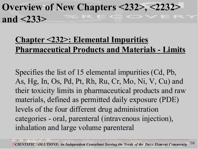
USP Chapter ⟨232⟩

USP very recently announced(4) a series of revisions to USP Chapter ⟨232⟩, Elemental Impurities, the revisions made being intended to align the general chapter more closely to ICH Q3D. One of the most significant is the removal of the need to routinely screen for Class 1 metals as part of any analysis, the final sentence in the text outlined below being deleted.

If, by process monitoring and supply chain control, manufacturers can demonstrate compliance, then further testing may not be needed. When testing is done to demonstrate compliance, proceed as directed in Elemental Impurities—Procedures ⟨233⟩.

This is a welcome and important amendment; the previous requirement making little scientific sense, there being no actual evidence that Class 1 metals would be more prevalent, for example, where a platinum catalyst was used than in the absence of a catalyst. Such catalysts are not a source of class 1 metals.

Overall

Overall there are likely to be challenges/uncertainties associated with ICH Q3D leading up to and for some period after the effective date as the guideline beds in, but the crucial fact is that all of the evidence to date indicates it is unlikely that there will be a widespread impact caused by issues of excessive levels of any elemental impurity whereby effective control cannot be realized.
/////////ICH Q3D, Elemental Impurities, Procedures ⟨233⟩, Training Modules
Share

ICH M8 “Specification for Submission Formats for eCTD”

 regulatory  Comments Off on ICH M8 “Specification for Submission Formats for eCTD”
May 172016
 

This additional specification describes the way files should be constructed for inclusion in the eCTD.

Key Points:

  • It is not necessary to use a product from Adobe or from any specific company to produce PDF documents.
  • All ICH regional regulatory authorities are able to read and accept PDF files saved as PDF version 1.4 through 1.7, PDF/A-1, or PDF/A-2 compliant to ISO 32000-1:2008.
  • The size of a PDF file should not exceed 500MB.

  • Regulatory authorities cannot guarantee the availability of any fonts except Times New Roman, Arial, and Courier and fonts supported in the Acrobat product set itself. Therefore, all additional fonts used in the PDF files should be embedded to ensure that those fonts would always be available to the reviewer.
  • Times New Roman, 12-point font, is adequate in size for narrative text and should be used whenever possible. Times New Roman font sizes 9-10 or an equivalent size of other recommended fonts are considered acceptable in tables but smaller font sizes should be avoided.
  • The use of a black font color is recommended. Blue can be used for hypertext links. Light colors can be difficult to read on a monitor as well as when printed, and should be avoided. The use of background shadowing can be difficult to read and should be avoided.
  • Pages should be properly oriented so that all portrait pages are presented in portrait and all landscape pages are presented in landscape.
  • A sufficient margin of at least 2.0 cm on the left side of each page for portrait and top of the page for landscape should be provided to avoid obscuring information. The remaining margins should be a Page 6 of 9minimum of 0.8 cm. Header and footer information can appear within these margins but should not appear so close to the page edge to risk being lost upon printing.
  • All pages of a document should include a unique header or footer that briefly identifies its subject matter.
  • Scanning should be avoided where possible.
  • It is recommended that scanning be undertaken at a resolution of 300 dots per inch (dpi) to balance legibility and file size. The use of grayscale or color is discouraged because of file size. After scanning, resampling to a lower resolution should be avoided.
  • Paper documents containing hand-written notes should be scanned at a resolution of at least 300 dpi. Hand-written notes should be done in black ink for clarity, 600 dpi is recommended. High-pressure liquid chromatography or similar images should be scanned at a resolution of at least 300 dpi.

  • Applicants should validate the quality of the renditions.
  • Hypertext links can be designated by rectangles using thin lines or by blue text as appropriate. Bookmarks are expected even if there is no TOC In the document. The use of no more than 4 levels in the hierarchy is recommended, but additional levels could be created for study reports if such bookmarks contribute to efficient navigation.
  • Relative paths should be used when creating hypertext links to minimize the loss of hyperlink functionality when folders are moved between disk drives.

  • The bookmarks should be collapsed when document is opened so that all bookmarks are at the first level.
  • The first page of the document should be numbered page 1, and all subsequent pages (including appendices and attachments) should be numbered consecutively with Arabic numerals. Roman numerals should not be used to number page. The only exception should be where a document is split because of its size, the second or subsequent file should be numbered consecutively to that of the first or preceding file.
  • Security fields should be set to allow printing, changes to the document, selecting text and graphics, and adding or changing notes and form fields. The exception to this rule includes regulatory forms with pre-existing security and literature references that need to be copyright protected.


Reference

http://estri.ich.org/ssf/Specification_for_Submission_Formats_for_eCTD_v1_0_.pdf

////////ICH M8, Specification, Submission Formats,  eCTD

Share

Ethyl 1-(7-chloroquinolin-4-yl)-5-methyl-1H-1,2,3-triazole-4-carboxylate

 Uncategorized  Comments Off on Ethyl 1-(7-chloroquinolin-4-yl)-5-methyl-1H-1,2,3-triazole-4-carboxylate
May 172016
 

STR1

STR1


Scheme 1 General scheme of the reaction. 

Scheme 2 

General procedure for the synthesis of 7-chloroquinoline-1,2,3-triazoyl carboxylates

To a solution of 4-azido-7-chloroquinoline 1 (0.3 mmol, 0.061 g) in DMSO (0.3 mL), was firstly added the β-ketoesters 2a-k (0.3 mmol) and then the catalyst pyrrolidine (0.03 mmol. 0.021 g). The reaction mixture was stirred in an open vial at room temperature for 24 hours. After completion of the reaction, the crude product was purified by column chromatography on silica gel using a mixture of hexanes/ethyl acetate (5:1) as the eluent to afford the desired products 3a-k.

Ethyl 1-(7-chloroquinolin-4-yl)-5-methyl-1H-1,2,3-triazole-4-carboxylate (3a)

Yield: 0.085 g (90%); white solid; mp 128-130 °C;

1H NMR (CDCl3, 300 MHz) δ 9.15 (d, 1H, J4.5 Hz, HetAr-H), 8.27 (d, 1H, J1.9 Hz, HetAr-H), 7.60 (dd, 1H, J9.0 and 1.9 Hz, HetAr-H), 7.48 (d, 1H, J4.5 Hz, HetAr-H), 7.34 (d, 1H, J9.0 Hz, HetAr-H), 4.50 (qua, 2H, J7.1 Hz, OCH2), 2.49 (s, 3H, CH3), 1.47 (t, 3H, J7.1 Hz, CH3);

13C NMR (CDCl3, 75 MHz) δ 161.10, 151.28, 149.88, 140.20, 139.34, 137.00, 136.76, 129.67, 128.93, 123.58, 122.09, 118.75, 61.15, 14.18, 9.44;

MS m/z (relative intensity): 316 (7), 259 (15), 243 (17), 231 (19), 217 (45), 215 (100), 214 (22), 205 (16), 203 (19), 189 (28), 181 (27), 179 (27), 164 (26), 162 (80), 137 (15), 135 (44), 127 (44), 126 (27), 100 (20), 99 (65), 83 (30), 75 (15), 74 (14), 43 (25);

HRMS calcd. for C15H14ClN4O2 [M + H]+: 317.0805; found: 317.0788.

Journal of the Brazilian Chemical Society

On-line version ISSN 1678-4790

J. Braz. Chem. Soc. vol.27 no.1 São Paulo Jan. 2016

http://dx.doi.org/10.5935/0103-5053.20150239

ARTICLES

7-Chloroquinoline-1,2,3-triazoyl Carboxylates: Organocatalytic Synthesis and Antioxidant Properties

Maiara T. Saraivaa  , Roberta Krügera  , Rodolfo S. M. Baldinottib  , Eder J. Lenardãoa  , Cristiane Lucheseb  , Lucielli Savegnagob  , Ethel A. Wilhelmb  *  , Diego Alvesa  * 

aLaboratório de Síntese Orgânica Limpa (LASOL, CCQFA), Universidade Federal de Pelotas (UFPel), CP 354, 96010-900 Pelotas-RS, Brazil

bGrupo de Pesquisa em Neurobiotecnologia (GPN), CDTec/CCQFA, Universidade Federal de Pelotas (UFPel), CP 354, 96010-900 Pelotas-RS, Brazil

ABSTRACT

We describe herein our results on the synthesis and antioxidant properties of 7-chloroquinoline-1,2,3-triazoyl-4-carboxylates. This class of compounds have been synthesized in moderated to excellent yields by the reaction of 4-azido-7-chloroquinoline with a range of β-ketoesters in the presence of a catalytic amount of pyrrolidine (10 mol%). The synthesized compounds ethyl 1-(7-chloroquinolin-4-yl)-5-methyl-1H-1,2,3-triazole-4-carboxylate and ethyl 1-(7-chloroquinolin-4-yl)-5-phenyl-1H-1,2,3-triazole-4-carboxylate were screened for their in vitro antioxidant activity and the results demonstrated that the first compound reduces the lipid peroxidation levels induced by sodium nitroprusside in liver of mice, while the second compound shown nitric oxide scavenging activity. This is an efficient method to produce new heterocyclic compounds with potential antioxidant activities.

http://www.scielo.br/scielo.php?script=sci_arttext&pid=S0103-50532016000100041&lng=en&nrm=iso&tlng=en

e-mail: diego.alves@ufpel.edu.br, ethelwilhelm@yahoo.com.br

Figure 1 Biologically important quinolines. 

Key words: quinolines,  1,2,3-triazoles,  organocatalysis,  cycloaddition,  antioxidant

/////////////

Share
Follow

Get every new post on this blog delivered to your Inbox.

Join other followers: